 |
 |
| Plant Pathol J > Volume 39(3); 2023 > Article |
|
Abstract
Citrus black rot is a serious disease of citrus plants caused by Alternaria citri. The current study aimed to synthesize zinc oxide nanoparticles (ZnO-NPs) by chemically or green method and investigate their antifungal activity against A. citri. The sizes of synthesized as measured by transmission electron microscope of ZnO-NPs were 88 and 65 nm for chemical and green methods, respectively. The studied prepared ZnO-NPs were applied, in vitro and in situ, at different concentrations (500, 1,000, and 2,000 μg/ml) in post-harvest treatment on navel orange fruits to verify the possible control effect against A. citri. Results of in vitro assay demonstrated that, at concentration 2,000 μg/ml, the green ZnO-NPs was able to inhibit about 61% of the fungal growth followed by 52% of chemical ZnO-NPs. In addition, scanning electron microscopy of A. citri treated in vitro with green ZnO-NPs showed swelling and deformation of conidia. Results showed also that, using a chemically and green ZnO-NPs at 2,000 μg/ml in situ in post-harvest treatment of orange, artificially-infected with A. citri, has reduced the disease severity to 6.92% and 9.23%, respectively, compared to 23.84% of positive control (non-treated fruits) after 20 days of storage. The out findings of this study may contribute to the development of a natural, effective, and eco-friendly strategy for eradicating harmful phytopathogenic fungi.
Citrus fruits are produced worldwide in tropical and subtropical areas and are the largest fruit commodities in international trade (FAOSTAT, 2018). After harvest, citrus fruits are susceptible to physiological disorders and microbial diseases that result in post-harvest losses. Furthermore, post-harvest citrus decay is the most serious cause of wastage and quality deterioration because it renders fresh fruit unfit for consumption, resulting in significant economic losses (Cheng et al., 2020; Garganese et al., 2019; Khorram and Ramezanian, 2021). In less developed countries, losses can reach 30% of total production and even 50% (Strano et al., 2022).
Some fruit post-harvest decays can be caused by latent infections in farms, which typically include infections caused by Alternaria alternata pv. citri Whiteside, and Phytophthora citrophthora, the causal agents of black and brown rots (Strano et al., 2022). The causal agent of citrus black rot is Alternaria citri, which is the first citrus-associated Alternaria species (Peever et al., 2004). Black rot of fruit, caused by A. citri, is a significant fungal disease of citrus that causes severe losses in citrus orchards of the world. It affects most of the members of the citrus family and often causes severe losses. The disease primarily affects plant above-ground parts, particularly leaves and fruits. The first symptoms, however, usually appear on the leaves as black necrotic lesions. Later, black necrotic lesions form on the fruits, and as the plants’ age, the fruits become soft and black-rotted (Umer et al., 2021). In a 10-year survey of citrus-associated diseases from several countries in the Mediterranean region, high disease pressure has been detected in all areas, forcing citrus growers to adapt fungicidal spray programs.
In fact, chemical control management poses serious risks to human health and the environment; additionally, continuous fungicide applications may result in the emergence of resistance to pathogenic species (Sánchez-Torres and Tuset, 2011; Vitale et al., 2021). Thus, there is a great interest to search for natural antifungal agents to control plant disease (Elshafie et al., 2017a, 2017b; Yang et al., 2019). Although these techniques are costly and time-consuming, it is critical to extend the technical life of fungicides after a certain period (Piccirillo et al., 2018). Furthermore, consumers are worried about the utilization of chemical fungicides since their active ingredients and co-formulants have been related to health problems and ecological contamination (Nicolopoulou-Stamati et al., 2016). Moringa oleifera Lam. (moringa) is a multi-purpose herbal plant used as human food and as an alternative for medicinal purposes worldwide. It has been identified by researchers as a plant with numerous health benefits, including nutritional and medicinal advantages (Abdull Razis et al., 2014).
Nanomaterials have been proposed and accepted due to their properties and critical applications in food and agriculture (Khan et al., 2019). In the field of post-harvest technology of fruits and vegetables, the nanotechnology approach is helpful for controlling post-harvest diseases, introducing an invention for packaging films, preventing the impact of gases and unsafe rays, improving packaging appearance, and helping to label fresh products using multiple chips (nano biosensors). Nanotechnology was used to produce nanomaterials for use as antifungal agents in several commodities, including fruits and vegetables (Ruffo Roberto et al., 2019). Due to their antibacterial properties, zinc oxide nanoparticles (ZnO-NPs) have received substantial attention (Meruvu et al., 2011). Even at low concentrations, the ZnO-NPs exhibited antibacterial and antifungal activities, making them acceptable for their coating applications (Sharma et al., 2010).
The present study used green-synthesized ZnO-NPs of M. oleifera as an alternative to toxic fungicides, in order to investigate ZnO-NPs’ antifungal effects against the A. citri Ellis & N. Pierce in vitro and in situ compared to the chemically-synthesized one. The study’s findings may help develop a new, efficient approach for controlling phytopathogenic fungi.
Zinc nitrate hexahydrate, sodium borohydride, and sodium hydroxide were obtained from Merck (KGaA, Darmstadt, Germany). Switch 62.5WG Fungicide was obtained from Syngenta USA (Greensboro, NC, USA).
The ZnO-NPs were prepared following the Wet-chemical method as reported by Arora et al. (2014). In particular, zinc nitrate and sodium hydroxide have been used as precursors, whereas sodium borohydride has been used as a stabilizing agent. Following the reaction and the formation of zinc hydroxide, calcinations should be performed to obtain the required size. Briefly, 2.79 g of Zn(NO3)2·6H2O was weighed and dissolved in 100 ml of distilled, deionized water, which was then agitated vigorously for 1 h. Then 0.48 g NaOH dissolved in 60 ml distilled deionized water was prepared, and then 1 ml NaBH4 (1%) was added dropwise after zinc nitrate dissolution, and stirred overnight for 12 h to obtain a low nano size. The precipitate was then allowed to settle before being filtered under suction and washed several times with distilled, deionized water (pump). The precipitate was then dried in an oven at 80°C. Zn(OH)2 was formed and then converted to ZnO. The muffle calcination had been performed at 500°C to obtain the smaller size.
The green synthesis of ZnO-NPs has been carried out using M. oleifera as follows. Ten grams of M. oleifera were obtained from the greenhouse of the Desert Research Center (Cairo, Egypt), washed with distilled water, and bot leaves surfaces were sterilized using alcohol by gentle rubbing. These leaves were heated for 40 min in 100 ml of distilled water at 50°C. The extract was filtered with Whatman 41 (Merck, KGaA). The filtrate was stored in a cool and dry place.
Zinc oxide nanoparticles were prepared by mixing 10 ml of M. oleifera leaf extract with 90 ml of a 0.05 mM zinc nitrate hexahydrate [Zn(NO3)2·6H2O] solution. The mixture was heated on a hot plate at 80°C for 30 min with continuous stirring. The presence of white-colored particles indicated the formation of nanoparticles. The extract was centrifuged and the pellets were collected, dried, and calcinated for 2 h at 300°C. White-colored nanoparticles were obtained as per the protocol as follows (El-Saber, 2021; Pal et al., 2018; Zambri et al., 2019).
The shape and size of ZnO-NPs were practically obtained using high-resolution transmission electron microscopy (TEM; HRTEM, JEOL JEM-2100, JEOL, Tokyo, Japan). Specimens for TEM measurements were prepared by placing a drop of colloidal solution on a 400-mesh carbon-coated copper grid and evaporating the solvent in the air at room temperature. The programme (ImageJ, U.S. National Institutes of Health, Bethesda, MD, USA) was used to calculate the average size of nanoparticles. The crystalline structure of the ZnO-NPs was confirmed via X-ray diffractometer (XRD; X‘Pert Pro, PanAlytical, Almelo, The Netherlands). Malvern zeta sizer nano was used to measure particle size and zeta potential. The sample in solution form was placed in a cuvette and mitigated to become transparent until the error in the reading was reduced, then was placed inside the device, and the device start required the diameter of the particles and determination of the extent of homogeneity through the polydispersity index (PDI; dispersed index), which ranges from 0 to 1, and when approached zero increased homogeneity.
Samples with visible disease symptoms were removed by a sterilized knife in such a manner as to contain the lesion edges too. These were then immersed in HgCl2 (1:10,000) for 2-3 min for surface sterilization and washed thoroughly in sterile distilled water until all the HgCl2 was removed. The surface-sterilized tissues were further cut into smaller pieces using a sterile blade and placed on potato dextrose agar (PDA) medium using disinfected forceps. Plates were incubated at 28°C for 7 days. The isolated fungi were identified by molecular methods based on polymerase chain reaction (PCR) (Camele et al., 2005) at Laboratory of Mycology, School of Agricultural, Forestry, Food and Environmental Sciences (SAFE), University of Basilicata (Potenza, Italy). The genomic DNA (gDNA) were extracted from each pure fungal isolate using DNeasy Plant Mini Kit (Qiagen, Heidelberg, Germany). The gDNAs were amplified using the universal primer pair ITS4/ITS5 of rDNA (White et al., 1990). The obtained amplicons were directly sequenced and the obtained sequences were compared with those available in the GenBank nucleotide archive using Basic Local Alignment Search Tool software BLAST (Bethesda, MD, USA) (Altschul et al., 1997). The purified isolates were maintained on PDA slants for further studies (Talibi et al., 2014).
The poisoned food in vitro technique described by Das et al. (2010) was employed to evaluate the antifungal activity of the tested materials (moringa leaves aqueous extract, ZnO-NPs, and green zinc nanoparticles) against A. citri at different concentrations (0, 500, 1,000, and 2,000 μg/ml) by incorporating appropriate quantities of the treatments with an autoclaved PDA at 45°C, mixing very well, then plating in sterile Petri dishes and allowing to solidify (Elshafie et al., 2020a, 2020b, 2021). With the assistance of a sterile cork borer, a 5 mm mycelial disc of the old culture of A. citri was taken and aseptically placed in the center of the treated plates. The inoculated plates were incubated at 28°C for 7 days, or until the control plate grew to 9 cm. The inhibition percentage was measured with the help of the mean colony diameter and computed according to the following formula (O’Rourke et al., 2009).
Where, C = Average mycelial growth in control petri plates, and T = Average mycelial growth in treated Petri plates.
Scanning electron microscopy (SEM) of A. citri treated with studied antifungal agents at 2,000 μg/ml compared to control (without treatment) for 4 h at room temperature was estimated according to Sitohy et al. (2013) as described by Abbas et al. (2020).
An in situ experiment has been carried out using navel orange fruit obtained from the local market. Harvested orange fruits were immediately transported to the Plant Pathology Department, Faculty of Agriculture, Zagazig University, Zagazig, Egypt some fruits were selected based on uniformity of size and color, then were disinfected using 70% ethanol for 1 min, 1% sodium hypochlorite for 1 min, and washed three times with sterilized distilled water, then drained, rinsed with fresh air, and air dried at room temperature (25°C) before being treated on the same day. A fresh PDA culture of A. citri, was prepared and used for artificial infection of navel orange fruits (six groups of six fruits each, in three replicates). Twenty microliters (2,000 μg/ml) of each tested material (moringa leaves aqueous extract, ZnO-NPs, and green zinc nanoparticles) were injected into the central axis of a sterilized fruit using a micropipette. Switch fungicide was similarly applied using the recommended dose as positive control. The negative control was treated likewise, except that the fruits were neither infected with the fungus nor treated with nanoparticles. After that, 100 μl of spore suspension containing 106 spores/ml was injected directly into the central axis of a sterilized fruit following the method described by Katoh et al. (2006). The inoculated fruits were then incubated in a sealed, sterilized plastic box at 24°C for 3 weeks, and the fruits were sliced in half for observation of symptom development. Disease severity was determined according to the area of the lesion on each fruit using the following formula:
The PCR amplification of the isolated fungi produced amplicons with molecular weights of approximately 600 bp. No amplification was observed in the case of the negative control. The obtained amplicons were directly sequenced (BMR Genomics, Padova, Italy) and the obtained sequences were compared with those available in the GenBank nucleotide archive using the Basic Local Alignment Search Tool software (BLAST) (Altschul et al., 1997). The sequences analysis for the isolated fungi showed high similarity with the sequences already presented in the GenBank database >99% for A. citri.
The chemical characterization of ZnO-NPs showed a well-dispersed, nearly spherical shape with a particle size of approximately 80 ± 5 nm as shown in the TEM image (Fig. 1A) and for extra-confirmatory identification by dynamic light scattering, which was performed to evaluate the nano-dispersity and particle size determination, an average particle size of 80 ± 5 nm with 0.552 PDI indicated a narrow size range (Fig. 1B). Stability and surface charge were estimated by the zeta potential exhibited by the −11.5 mV moderately stable NPs (Fig. 1C). For phase analysis and chemical structural identification, the XRD technique was conducted, where the XRD pattern of ZnO-NPs showed peaks at 2 = 31.69, 34.39, 36.21, 47.51, 56.47, 62.97, and 68.13, card number 01-078-2585 hexagonal (Fig. 1D).
Different characterization techniques including TEM, XRD, zeta potential, and zeta size were used to ascertain the formation of ZnO-NPs green synthesized by moringa leaf. The shape and the size of the biosynthesized ZnO-NPs were studied and investigated by using TEM (Fig. 2A). The average size and the stability of the nanoparticles were confirmed by the zeta size (Fig. 2B) and the zeta potential analysis (Fig. 2C). The obtained results showed that the mean diameter of ZnO-NPs was about 65 nm, and the zeta potential was −13.7 mv. Finally, The XRD pattern of the ZnO-NPs powder showed various diffractions peaks at 2 theta degree 31.24, 34.11, 35.71, 47.77, 56.22, 62.72, 66.34, 67.78, 72.84, and 76.79. As matched with the crystallographic database JCPDS 36-1451, these peaks were assigned to (100), (002), (101), (102), (110), (103), (200), (112), (004), and (202) planes of zinc, respectively (Fig. 2D).
Figs. 3 and 4 illustrate the in vitro antifungal activity of M. oleifera leaf aqueous extract and ZnO-NPs prepared either chemically or green-synthesis against A. citri mycelium growth. All treatments showed a promising effect on the linear growth of A. citri in a dose-dependent manner compared to control. A linear growth percentage reduction of A. citri at a concentration was 2,000 μg/ml, which was the most effective, being 3.5, 4.36, and 5.3 cm for greenly synthesized ZnO-NPs, chemically-synthesized ZnO-NPs, and moringa aqueous extract, respectively. Furthermore, data showed that the green-synthesized ZnO-NPs at 2,000 μg/ml was the most effective dose decreasing the linear growth of A. citri, where it showed 61.1%, followed by chemical NPs (51.55%), and finally moringa aqueous leaf extract (41.11%).
SEM was used to examine the morphological features of A. citri mycelia and cells treated with green-synthesized ZnO-NPs at 2,000 μg/ml to investigate the possible antifungal mechanism of these NPs against A. citri. The observation explicated that the mycelia and conidia of A. citri grown on PDA were smooth, homogeneous mycelial morphology. In contrast, the mycelial and conidial morphology was conspicuously changed when exposed to green-synthesized ZnO-NPs at 2,000 μg/ml (Fig. 5).
Table 1 and Fig. 6 showed the effect of moringa leaf aqueous extract and new green and chemically-synthesized ZnO-NPs on the severity of black rot disease on navel orange. Data presented in Table 1 indicates that switch fungicide completely inhibited black rot disease severity. Furthermore, data showed that green ZnO-NPs at 2,000 μg/ml was the most effective in reducing black rot disease severity (93.08%), followed by chemical ZnO-NPs (90.77%), whereas moringa aqueous extract exhibited only 76.93% effectiveness.
Citrus fruit rot disease is incited by A. citri is one of the serious diseases that attack citrus cultivars, causing severe damage and causing many diseases, i.e., black fruit rot, stem end rot, brown spot, and leaf spot (Isshiki et al., 2001; Sadowsky et al., 2002). Citrus black rot is a major fungal disease of sweet oranges (Citrus sinensis L.) that affects the fruit even after post-harvest, leading to a devastating impact on the overall production of oranges. A. citri, the prime etiological agent of the disease, has been reported to develop fungicide-resistance during the last decade (Sardar et al., 2022; Zelmat et al., 2021). Hence, there is a need to use alternative techniques to control fungal infections. Nanoparticles can be used as possible alternative to control phytopathogens in comparison to synthetic fungicides. ZnO-NPs have wider applications and are generally recognized as safe materials by the US Food and Drug Administration (Isshiki et al., 2001). The application of ZnO-NPs has been recently increased including cosmetics ointments, food packaging, etc. (Bobo et al., 2016; Espitia et al., 2012; Marcous et al., 2017; Pillai et al., 2020). Pillai et al. (2020) reported that the antifungal activity of ZnO-NPs was prepared by the green method, and found that ZnO-NPs prepared using Beta vulgaris were active against Aspergillus niger, while those prepared from Cinnamomum tamala were active against Candida albicans. Additionally, ZnO-NPs were prepared from the extract of Brassica oleracea var. italica has shown activity against both fungal strains.
Antifungal activity of ZnO-NPs (35-45 nm), silver (20-80 nm), and titanium dioxide (85-100 nm) has been tested against Macrophomina phaseolina, a major soil-borne pathogen of pulse and oilseed crops. Silver nanoparticles (AgNO3 NPs) had a greater antifungal effect at lower concentration than ZnO-NPs and TiO2 NPs. Maize treated with nanosilica (20-40 nm) has been screened for resistance against Fusarium oxysporum and A. niger compared to that of bulk silica (Shyla et al., 2014).
The normal mycelial and conidial morphology changed noticeably when exposed to green ZnO-NPs at 2,000 μg/ml in the current study. The mode of action of prepared NPs may be explained through the electrostatic interaction between metal ions and the microbial cell membrane, resulting in damage to the cell membrane and intracellular organelles (Nisar et al., 2019). These continued steps enhance the inhibitory activity of NPs (Gold et al., 2018). The penetration of NPs into microbial cells increases the microbial inhibition interacting with the electron transport chain, disrupting DNA by breaking phosphate and hydrogen bonds, denaturation proteins by altering tertiary structure, and killing mitochondria via oxidative stress (Stanić and Tanasković, 2020). Due to the interactions between inorganic metal and metal oxide NPs, reactive oxygen species are produced causing cell damage (Abd-Ellatif et al., 2022; Canaparo et al., 2020). Greenly manufactured inorganic metal and metal oxide nanoparticles have a wide range of applications in agriculture, particularly in the management of fungal plant diseases (Abd-Ellatif et al., 2022).
The usage of green NPs as potential bio-pesticides should replace the detrimental effects of conventional pesticides for a sustainable environment. One of the most prevalent post-harvest diseases affecting navel oranges is citrus black rot, which has a significant negative influence on citrus fruit worldwide. The diseases resistance which frequently occurred in case of conventional fungicides draws the researchers’ attention to find out novel, effective natural alternatives. Nanoparticles have been reported to have a good long-term antifungal effect. Green-synthesized ZnO-NPs showed a greater antifungal efficacy than the chemically synthesized one against A. citri, the causal agent of citrus black rot disease. In conclusion, the use of eco-friendly green-synthesized metal oxide NPs may reduce the fungal infection of navel orange and improve the farmers’ socio-economic standings.
Acknowledgments
This research was funded by the project entitled “Fighting plant fungi post-harvest using environmentally friendly bio products’, financially supported by the Science and Technology Development Fund (STDF), Egypt. Grant type/name: Research Support. Project ID: 43673.
Fig. 1
Transmission electron microscopy (A), particle size (B), zeta potential (C), and X-ray diffractometer (XRD) pattern (D) zinc oxide nanoparticles prepared using chemical method.
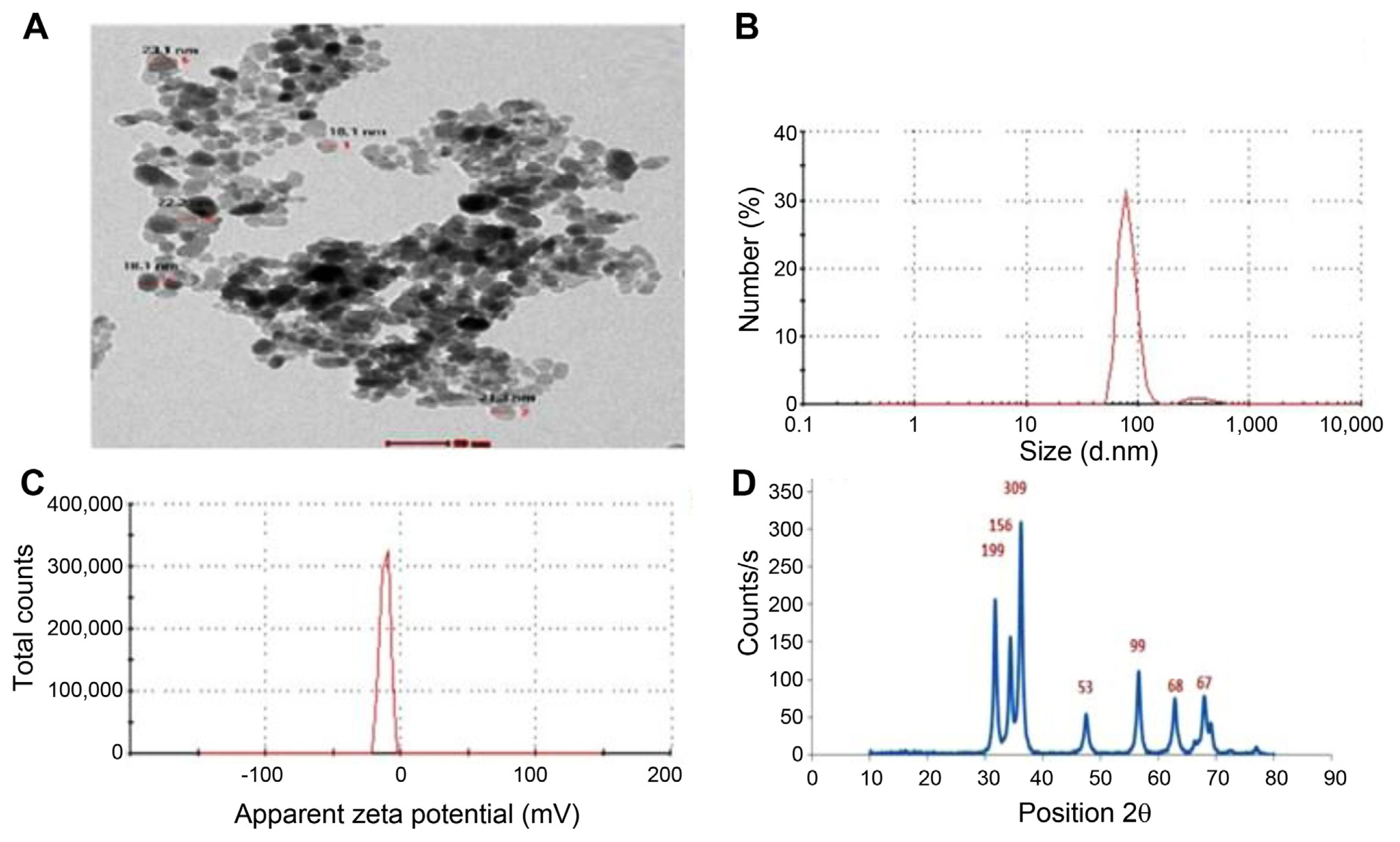
Fig. 2
Transmission electron microscopy (A), particle size (B), zeta potential (C), and X-ray diffractometer (XRD) pattern (D) of zinc oxide nanoparticles prepared using green method.
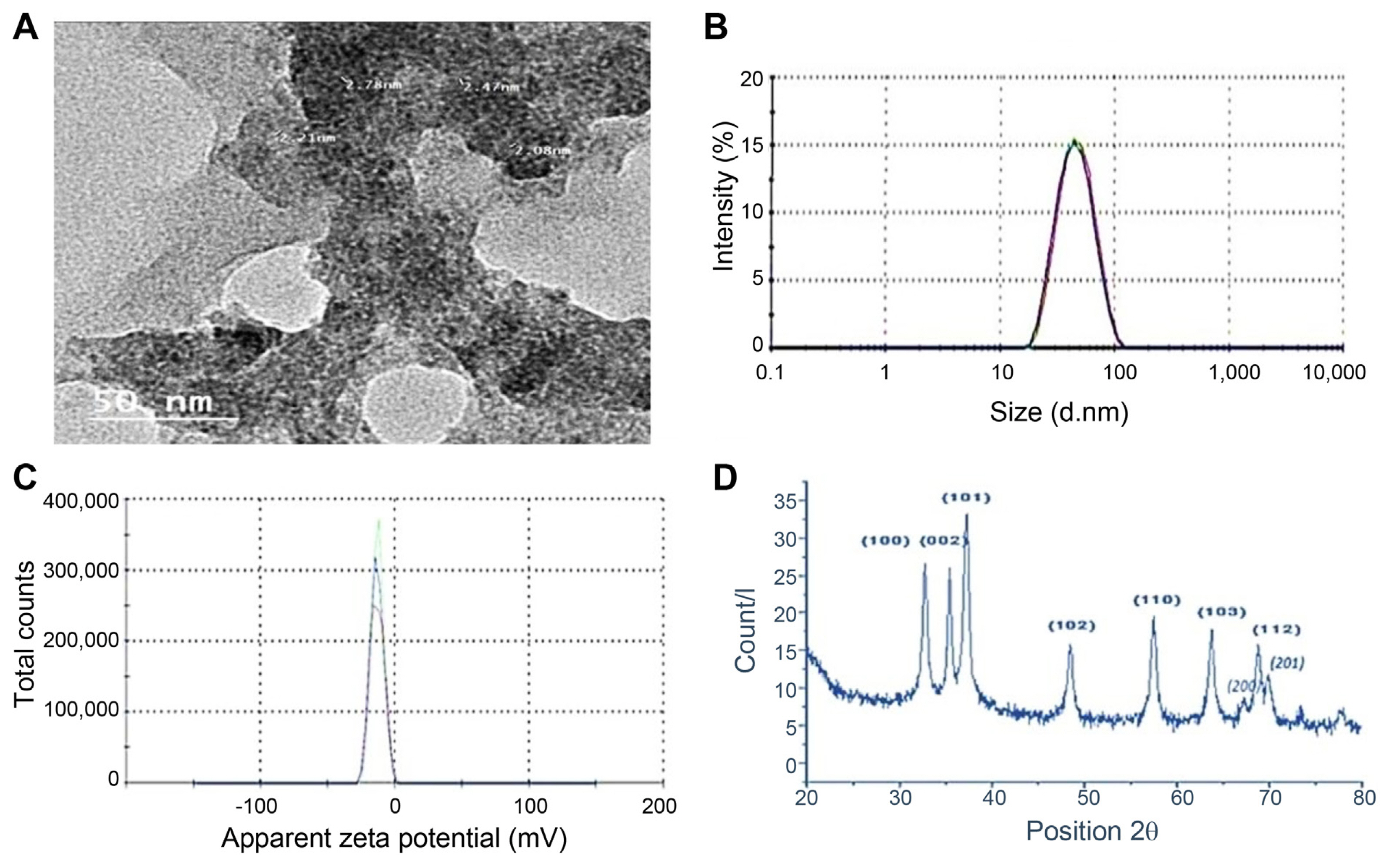
Fig. 3
Fungal growth of Alternaria citri on potato dextrose agar medium after incubation at 28°C for 7 days with Moringa oleifera leaf aqueous extract (A), chemical zinc oxide nanoparticles (ZnO-NPs) (B), and green ZnO-NPs (C) at different concentrations (500, 1,000, and 2,000 μg/ml) compared to control (non-treated plates).
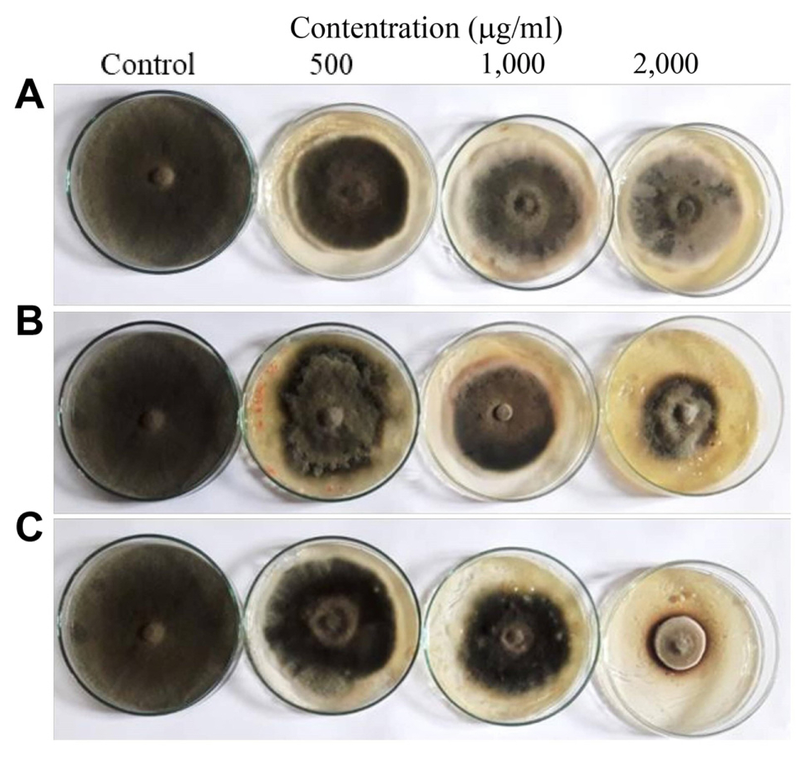
Fig. 4
Linear growth (cm) (A) and growth reduction percent (B) of Alternaria citri on potato dextrose agar medium after 7 days of incubation at 28°C with Moringa oleifera leaf aqueous extract, chemical zinc oxide nanoparticles (ZnO-NPs), green ZnO-NPs at different concentrations (500, 1,000, and 2,000 μg/ml) compared to control (non-treated plates).
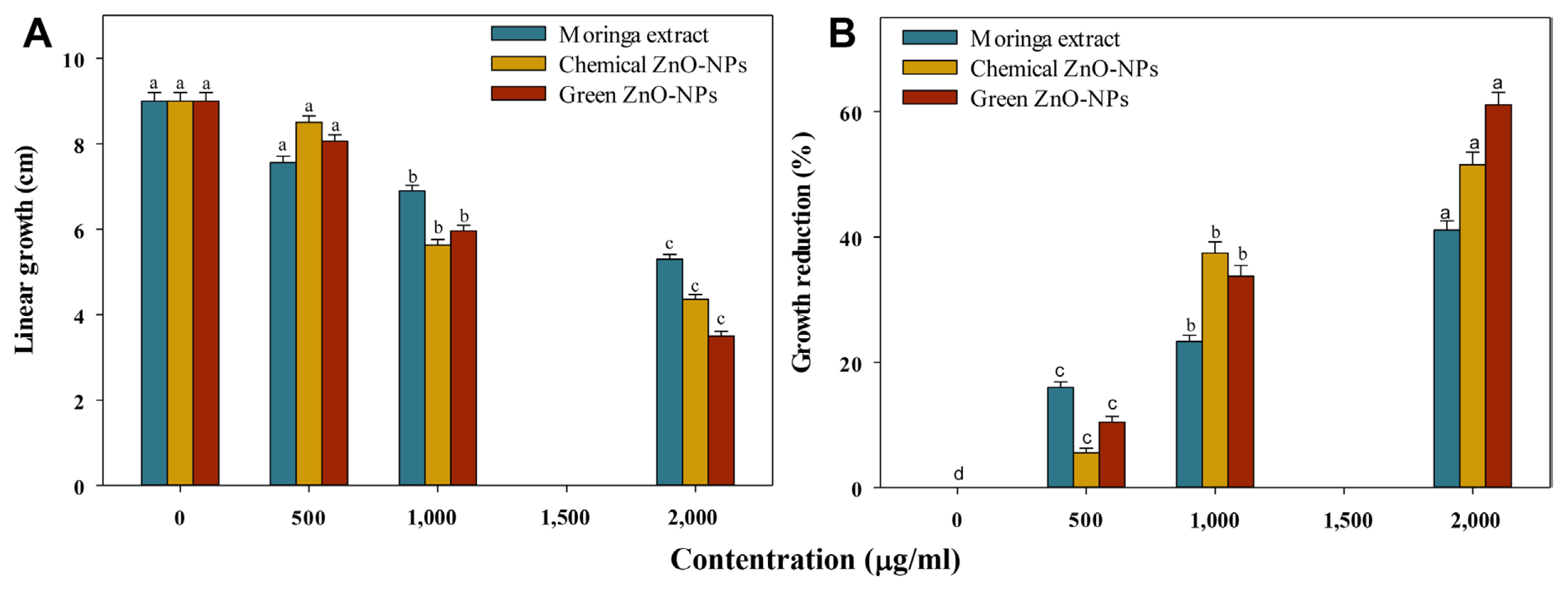
Fig. 5
(A-D) Scanning electron microscopy images of green-synthesized zinc oxide nanoparticles (ZnO-NPs; 2,000 μg/ml) against Alternaria citri. Arrows refer to the changes that were observed in the control and treatment.

Fig. 6
Different treatments of navel orange fruits infected with Alternaria citri after 3 weeks of incubation at 25 ± 2°C. ZnO-NP, zinc oxide nanoparticle.
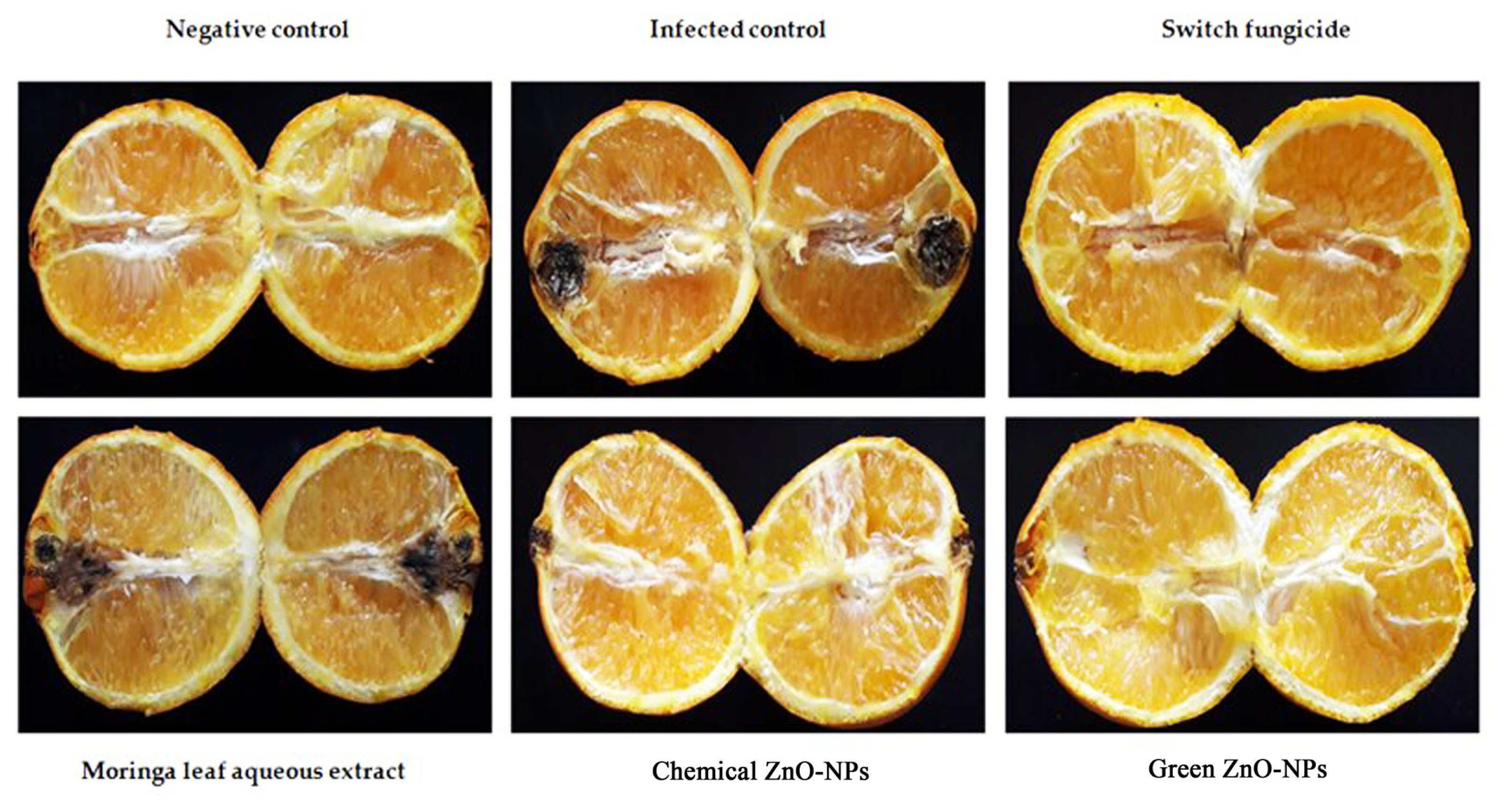
Table 1
Different treatments affecting navel orange black rot disease (in situ trial)
References
Abbas, E., Osman, A. and Sitohy, M. 2020. Biochemical control of Alternaria tenuissima infecting post-harvest fig fruit by chickpea vicilin. J. Sci. Food Agric 100:2889-2897.


Abd-Ellatif, S., Ibrahim, A. A., Safhi, F. A., Abdel Razik, E. S., Kabeil, S. S. A., Aloufi, S., Alyamani, A. A., Basuoni, M. M., ALshamrani, S. M and Elshafie, H. S 2022. Green synthesized of Thymus vulgaris chitosan nanoparticles induce relative WRKY-genes expression in Solanum lycopersicum against Fusarium solani, the causal agent of root rot disease. Plants 11:3129.


Abdull Razis, A. F., Ibrahim, M. D and Kntayya, S. B 2014. Health benefits of Moringa oleifera. Asian Pac. J. Cancer Prev 15:8571-8576.


Altschul, S. F., Madden, T. L., Schäffer, A. A., Zhang, J., Zhang, Z., Miller, W. and Lipman, D. J 1997. Gapped BLAST and PSIBLAST: a new generation of protein database search programs. Nucleic Acids Res 25:3389-3402.


Arora, A. K., Devi, S., Jaswal, V. S., Singh, J., Kinger, M. and Gupta, V. D 2014. Synthesis and characterization of ZnO nanoparticles. Orient. J. Chem 30:1671-1679.

Bobo, D., Robinson, K. J., Islam, J., Thurecht, K. J and Corrie, S. R 2016. Nanoparticle-based medicines: a review of FDA-approved materials and clinical trials to date. Pharm. Res 33:2373-2387.


Camele, I., Marcone, C. and Cristinzio, G. 2005. Detection and identification of Phytophthora species in southern Italy by RFLP and sequence analysis of PCR-amplified nuclear ribosomal DNA. Eur. J. Plant Pathol 113:1-14.


Canaparo, R., Foglietta, F., Limongi, T. and Serpe, L. 2020. Biomedical applications of reactive oxygen species generation by metal nanoparticles. Materials 14:53.


Cheng, Y., Lin, Y., Cao, H. and Li, Z. 2020. Citrus postharvest green mold: recent advances in fungal pathogenicity and fruit resistance. Microorganisms 8:449.


Das, K., Tiwari, R. K. S and Shrivastava, D. K 2010. Techniques for evaluation of medicinal plant products as antimicrobial agent: current methods and future trends. J. Med. Plants Res 4:104-111.
El-Saber, M. M 2021. Effect of biosynthesized Zn and Se nanoparticles on the productivity and active constituents of garlic subjected to saline stress. Egypt. J. Desert Res 71:99-128.

Elshafie, H. S., Camele, I., Sofo, A., Mazzone, G., Caivano, M., Masi, S. and Caniani, D. 2020a. Mycoremediation effect of Trichoderma harzianum strain T22 combined with ozonation in diesel-contaminated sand. Chemosphere 252:126597.


Elshafie, H. S., Caputo, L., De Martino, L., Grul’ov́, D., Zheljazkov, V.Z., De Feo, V. and Camele, I. 2020b. Biological investigations of essential oils extracted from three Juniperus species and evaluation of their antimicrobial, antioxidant and cytotoxic activities. J. Appl. Microbiol 129:1261-1271.


Elshafie, H. S., Caputo, L., De Martino, L., Sakr, S. H., De Feo, V. and Camele, I. 2021. Study of bio-pharmaceutical and antimicrobial properties of pomegranate (Punica granatum L.) leathery exocarp extract. Plants 10:153.


Elshafie, H. S., Sakr, S., Bufo, S. A and Camele, I. 2017a. An attempt of biocontrol the tomato-wilt disease caused by Verticillium dahliae using Burkholderia gladioli pv. agaricicola and its bioactive secondary metabolites. Int. J. Plant Biol 8:7263.


Elshafie, H. S., Viggiani, L., Mostafa, M. S., El-Hashash, M. A., Camele, I. and Bufo, S. A 2017b. Biological activity and chemical identification of ornithine lipid produced by Burkholderia gladioli pv. agaricicola ICMP 11096 using LC-MS and NMR analyses. J. Biol. Res 90:6534.


Espitia, P. J. P., Soares, N. F. F., Coimbra, J. S. R., de Andrade, N. J., Cruz, R. S and Medeiros, E. A. A 2012. Zinc oxide nanoparticles: synthesis, antimicrobial activity and food packaging applications. Food Bioprocess Technol 5:1447-1464.


FAOSTAT. 2018 Statistical databases, fisheries data, 2001 Food and Agriculture Organization of the United Nations, Rome, Italy. URL https://www.fao.org/. 2 March 2023.
Garganese, F., Sanzani, S. M., Di Rella, D., Schena, L. and Ippolito, A. 2019. Pre- and postharvest application of alternative means to control Alternaria Brown spot of citrus. Crop Prot 121:73-79.

Gold, K., Slay, B., Knackstedt, M. and Gaharwar, A. K 2018. Antimicrobial activity of metal and metal-oxide based nanoparticles. Adv. Ther 1:1700033.


Isshiki, A., Akimitsu, K., Yamamoto, M. and Yamamoto, H. 2001. Endopolygalacturonase is essential for citrus black rot caused by Alternaria citri but not brown spot caused by Alternaria alternata. Mol. Plant-Microbe Interact 14:749-757.


Katoh, H., Isshiki, A., Masunaka, A., Yamamoto, H. and Akimitsu, K. 2006. A virulence-reducing mutation in the postharvest citrus pathogen Alternaria citri. Phytopathology 96:934-940.


Khan, I., Saeed, K. and Khan, I. 2019. Nanoparticles: properties, applications and toxicities. Arab. J. Chem 12:908-931.

Khorram, F. and Ramezanian, A. 2021. Cinnamon essential oil incorporated in shellac, a novel bio-product to maintain quality of ‘Thomson navel’ orange fruit. . J. Food Sci. Technol 58:2963-2972.


Marcous, A., Rasouli, S. and Ardestani, F. 2017. Low-density polyethylene films loaded by titanium dioxide and zinc oxide nanoparticles as a new active packaging system against Escherichia coli O157:H7 in fresh calf minced meat. Packag. Technol. Sci 30:693-701.


Meruvu, S., Hugendubler, L. and Mueller, E. 2011. Regulation of adipocyte differentiation by the zinc finger protein ZNF638. J. Biol. Chem 286:26516-26523.


Nicolopoulou-Stamati, P., Maipas, S., Kotampasi, C., Stamatis, P. and Hens, L. 2016. Chemical pesticides and human health: the urgent need for a new concept in agriculture. Front. Public Health 4:148.


Nisar, P., Ali, N., Rahman, L., Ali, M. and Shinwari, Z. K 2019. Antimicrobial activities of biologically synthesized metal nanoparticles: an insight into the mechanism of action. J. Biol. Inorg. Chem 24:929-941.


O’Rourke, TA, Scanlon, TT, Ryan, MH, Wade, LJ, McKay, AC, Riley, IT, Li, H, Sivasithamparam, K and Barbetti, MJ 2009. Severity of root rot in mature subterranean clover and associated fungal pathogens in the wheatbelt of Western Australia. Crop Pasture Sci 60:43-50.

Pal, S., Mondal, S., Maity, J. and Mukherjee, R. 2018. Synthesis and characterization of ZnO nanoparticles using Moringa oleifera leaf extract: investigation of photocatalytic and antibacterial activity. Int. J. Nanosci. Nanotechnol 14:111-119.
Peever, T. L., Su, G., Carpenter-Boggs, L. and Timmer, L. W 2004. Molecular systematics of citrus-associated Alternaria species. Mycologia 96:119-134.


Piccirillo, G., Carrieri, R., Polizzi, G., Azzaro, A., Lahoz, E., Fernández-Ortuño, D. and Vitale, A. 2018. In vitro and in vivo activity of QoI fungicides against Colletotrichum gloeosporioides causing fruit anthracnose in Citrus sinensis. Sci. Hortic 236:90-95.

Pillai, A. M., Sivasankarapillai, V. S., Rahdar, A., Joseph, J., Sadeghfar, F., Anuf, A. R., Rajesh, K. and Kyzas, G. Z 2020. Green synthesis and characterization of zinc oxide nanoparticles with antibacterial and antifungal activity. J. Mol. Struct 1211:128107.

Ruffo Roberto, S., Youssef, K., Hashim, A. F and Ippolito, A. 2019. Nanomaterials as alternative control means against postharvest diseases in fruit crops. Nanomaterials 9:1752.


Sadowsky, A., Kimchi, M., Oren, Y. and Solel, Z. 2002. Occurrence and management of Alternaria brown spot in Israel. Phytoparasitica 30:19.
Sánchez-Torres, P and Tuset, JJ 2011. Molecular insights into fungicide resistance in sensitive and resistant Penicillium digitatum strains infecting citrus. Postharvest Biol. Technol 59:159-165.

Sardar, M., Ahmed, W., Al Ayoubi, S., Nisa, S., Bibi, Y., Sabir, M., Khan, M. M., Ahmed, W. and Qayyum, A. 2022. Fungicidal synergistic effect of biogenically synthesized zinc oxide and copper oxide nanoparticles against Alternaria citri causing citrus black rot disease. Saudi J. Biol. Sci 29:88-95.


Sharma, D., Rajput, J., Kaith, B. S., Kaur, M. and Sharma, S. 2010. Synthesis of ZnO nanoparticles and study of their antibacterial and antifungal properties. Thin Solid Films 519:1224-1229.

Shyla, K. K., Natarajan, N. and Nakkeeran, S. 2014. Antifungal activity of zinc oxide, silver and titanium dioxide nanoparticles against Macrophomina phaseolina. J. Mycol. Plant Pathol 44:268-273.
Sitohy, M., Mahgoub, S., Osman, A., El-Masry, R. and Al-Gaby, A. 2013. Extent and mode of action of cationic legume proteins against Listeria monocytogenes and Salmonella enteritidis. Probiotics Antimicrob. Proteins 5:195-205.


Stanić, V. and Tanasković, S. B 2020. Antibacterial activity of metal oxide nanoparticles. In: Nanotoxicity: prevention and antibacterial applications of nanomaterials, eds. by S Rajendran, A Mukherjee, TA Nguyen, C Godugu and RK Shukla, pp. 241-274. Elsevier, Amsterdam, The Netherlands.
Strano, M. C., Altieri, G., Allegra, M., Di Renzo, G. C., Paterna, G., Matera, A. and Genovese, F. 2022. Postharvest technologies of fresh citrus fruit: advances and recent developments for the loss reduction during handling and storage. Horticulturae 8:612.

Talibi, I., Boubaker, H., Boudyach, E. H and Ait Ben Aoumar, A. 2014. Alternative methods for the control of postharvest citrus diseases. J. Appl. Microbiol 117:1-17.


Umer, M., Mubeen, M., Ateeq, M., Shad, M. A., Atiq, M. N., Kaleem, M. M., Iqbal, S., Shaikh, A. A., Ullah, I., Khan, M., Kalhoro, A. A and Abbas, A. 2021. Etiology, epidemiology and management of citrus black rot caused by Alternaria citri: an outlook. Plant Prot 5:105-115.


Vitale, A., Aiello, D., Azzaro, A., Guarnaccia, V. and Polizzi, G. 2021. An eleven-year survey on field disease susceptibility of citrus accessions to Colletotrichum and Alternaria species. Agriculture 11:536.

White, T. J., Bruns, T., Lee, S. and Taylor, J. 1990. Amplification and direct sequencing of fungal ribosomal RNA genes for phylogenetics. In: PCR protocols: a guide to methods and applications, eds. by MA Innis, DH Gelfand, JJ Sninsky and TJ White, pp. 315-322. Academic Press, New York, NY, USA.

Yang, L.-N., He, M.-H., Ouyang, H.-B., Zhu, W., Pan, Z.-C., Sui, Q.-J., Shang, L.-P and Zhan, J. 2019. Cross-resistance of the pathogenic fungus Alternaria alternata to fungicides with different modes of action. BMC Microbiol 19:205.


- TOOLS
-
METRICS

- ORCID iDs
-
Ali Osman

https://orcid.org/0000-0001-7174-0207Ippolito Camele

https://orcid.org/0000-0001-8099-8234 - Related articles


 PDF Links
 PubReader
 ePub Link
 Full text via DOI
 Full text via PMC
 Download Citation
 Print
Print


