Morphogenetic Alterations of Alternaria alternata Exposed to Dicarboximide Fungicide, Iprodione
Article information
Abstract
Fungicide-resistant Alternaria alternata impede the practical control of the Alternaria diseases in crop fields. This study aimed to investigate cytological fungicide resistance mechanisms of A. alternata against dicarboximide fungicide iprodione. A. alternata isolated from cactus brown spot was cultured on potato-dextrose agar (PDA) with or without iprodione, and the fungal cultures with different growth characteristics from no, initial and full growth were observed by light and electron microscopy. Mycelia began to grow from one day after incubation (DAI) and continued to be in full growth (control-growth, Con-G) on PDA without fungicide, while on PDA with iprodione, no fungal growth (iprodione-no growth, Ipr-N) occurred for the first 3 DAI, but once the initial growth (iprodione-initial growth, Ipr-I) began at 4–5 DAI, the colonies grew and expanded continuously to be in full growth (iprodione-growth, Ipr-G), suggesting Ipr-I may be a turning moment of the morphogenetic changes resisting fungicidal toxicity. Con-G formed multicellular conidia with cell walls and septa and intact dense cytoplasm. In Ipr-N, fungal sporulation was inhibited by forming mostly undeveloped unicellular conidia with degraded and necrotic cytoplasm. However, in Ipr-I, conspicuous cellular changes occurred during sporulation by forming multicellular conidia with double layered (thickened) cell walls and accumulation of proliferated lipid bodies in the conidial cytoplasm, which may inhibit the penetration of the fungicide into conidial cells, reducing fungicide-associated toxicity, and may be utilized as energy and nutritional sources, respectively, for the further fungal growth to form mature colonies as in Ipr-G that formed multicellular conidia with cell walls and intact cytoplasm with lipid bodies as in Con-G.
Introduction
The fungal genus Alternaria contains 299 species that are ubiquitous in the environment, where they act as human allergens for hay fever and asthma and/or as major plant pathogens (Nowicki et al., 2012; Pratt, 1941). Alternaria alternata is one of the most common species in this genus. It usually is a saprophyte, but also a necrotrophic plant pathogen on multiple host plants (Hutton and Mayers, 1988; Kohmoto et al., 1995; Nishimura and Kohmoto, 1983; Rotem, 1994). Chemical control with fungicides is commonly used to reduce Alternaria diseases both in fields and in storages of various economic plants (Filadić and Sutton, 1992; Humpherson-Jones and Maude, 1982; Hutton and Mayers, 1988; Lee and Kim, 1980; Maude and Humpherson-Jones, 2008; Maude et al., 1984; Miles et al., 2005; Singh et al., 2006; Swart et al., 1998; Tronchso-Rojas and Triznado-Hernadez, 2014; Tsedaley, 2014).
Dicarboximides are protectant fungicides that are effective against several fungal genera, including Alternaria, Botrytis, Sclerotinia, and Phoma (Steel and Nair, 1995). Iprodione was first developed and commercialized in 1974 (Lacroix et al., 1974), and is one of the best fungicides for controlling Alternaria blotch of apples (Lee and Kim, 1980; Sakurai and Fujita, 1978). However, iprodione-resistant Alternaria isolates are frequently found in crop fields and reduce the control of the Alternaria diseases (Dry et al., 2004; Hutton, 1988; McPhee, 1980; Solel et al., 1996).
Plant fungal pathogens can develop resistance to fungicides following continuous and widespread uses. This resistance can be prevented and delayed at least by using mixtures of and changing between fungicides with different modes of action during a cropping period (Deising et al., 2008). For this, the resistance mechanisms against the antifungal activities should be revealed to cope with the fungicide-resistant fungal strains by nullifying or attenuating the expression of their resistance mechanisms. For iprodione, the genetic and biochemical bases for this fungicide resistance have been documented (Dry et al., 2004; Steel and Nair, 1995). In Botrytis cinerea, most of the mutants resistant to iprodione showed modifications in the osmosensing class III histidine kinase affecting the HAMP domains (Fillinger et al., 2012). However, little has been documented on the cytological mechanisms of iprodione resistance in A. alternata, which may display microscopic alterations in phenotypic functions of cells and cellular organelles. The objectives of this study, therefore, are to reveal morphogenetic alterations of A. alternata in the fungal life cycle that are functionally related to the fungicide resistance. For this, an isolate of A. alternata was placed on potato-dextrose agar (PDA) amended with or without iprodione, and the portions of the fungal culture showing different growth characteristics from no, initial, and full growth were examined at different days of incubation by light microscopy (LM), scanning electron microscopy (SEM), and transmission electron microscopy (TEM).
Fungus, fungicide and the fungal growth
A. alternata CD2-7A isolated from the brown spot of a grafted cactus, Gymnocalycium mihanovichii (Choi et al., 2010) was used in the present study. The fungicide used was iprodione (50% wettable powder) [3-(3,5-dichlorophenyl)-A′-isopropyl-2,4-dioxoimidazolidine-l-carboximide] (Rovral®; Nonghyup Chemical, Seongnam, Korea). For the fungicidal test, the fungal isolate was grown on PDA at 25°C for 7 days. Fungal plugs were cut out from the fungal culture using a 5-mm-diameter corker borer and placed on PDA supplemented with 0.1% iprodione with 5 replications for each plate. PDA without fungicide was used as a control. These plates were incubated at 25°C under a 24-h illumination at a distance of 20 cm from a 40-W fluorescence lamp in an incubation chamber and the fungal growth was examined daily for 10 days after incubation (DAI). The experimentation was repeated on three different occasions.
Light and electron microscopy
Fungal specimens for light and electron microscopy were prepared from the 5-day-old cultures of A. alternata; one from the fully grown culture (control-growth, Con-G) in untreated control and three from no grown (dead) (iprodione-no growth, Ipr-N), initially growing (living) (iprodione-initial growth, Ipr-I), and fully grown (iprodione-growth, Ipr-G). Hyphae and conidia from the fungal cultures were observed under a compound light microscope (Axiophot; Zeiss, Oberkochen, Germany).
For electron microscopy, the fungal specimens were excised and fixed in Karnovsky’s fixative in 50 mM cacodylate buffer (pH 7.2) for 2 h (Karnovsky, 1965); rinsed in the same buffer solution three times each for 20 min. The fixed specimens were post-fixed in 1% osmium tetroxide in 50 mM cacodylate buffer for 2 h, washed briefly in distilled water and pre-stained in bulk overnight in 0.5% uranyl acetate at 4°C. The fixed specimens were then dehydrated in an ethanol series (30%, 50%, 80%, 95%, and three times of 100%) each for 10 min.
For SEM, the dehydrated specimens were dried in a transitional hydrophobic solvent (100% isoamylacetate) twice for 10 min each at room temperature and critical-point dried in an automated critical point dryer (EM CPD300; Leica Microsystems, Wetzlar, Germany). The dried specimens were coated with gold in a sputter coater (JFC-1110E; JEOL, Tokyo, Japan), and observed under a scanning electron microscope (JSM-5410LV; JEOL) at 20 kV.
For TEM, the dehydrated specimens from the ethanol series were further dehydrated in propylene oxide for 15 min, then placed in a 1:1 mixture of propylene oxide and Spurr’s epoxy resin for 5 h, and embedded in Spurr’s epoxy resin, followed by the polymerization of the resin at 70°C for 8 h (Spurr, 1969). The embedded specimens were sectioned with a diamond knife on an MT-X ultra-microtome (RMC Boeckeler, Tucson, AZ, USA) to make ultrathin sections of 80–90 nm thick. These sections were stained with 2% uranyl acetate and lead citrate for 7 min each and examined under a transmission electron microscope (JEM-1010; JEOL) at an acceleration voltage of 80 kV. At least three samples were observed per treatment replicate.
Fungal growth on PDA supplemented with or without iprodione
On the untreated control plate, the initially observed fungal growth was hyphal outgrowth from the inoculated agar plugs at one DAI (Fig. 1A). The fungal colonies expanded more after the initial growth, showing steady and uniform mycelial growths around the fungal colonies at two and three DAI (Fig. 1B, C). However, no fungal outgrowth occurred on PDA supplemented with iprodione until 3 DAI (Fig. 1D–F), on which the fungal outgrowth initiated at 4–5 DAI, followed by the occurrence of additional fungal outgrowths that expanded more outward with increased DAI (Fig. 1G–I).
Light microscopy and SEM
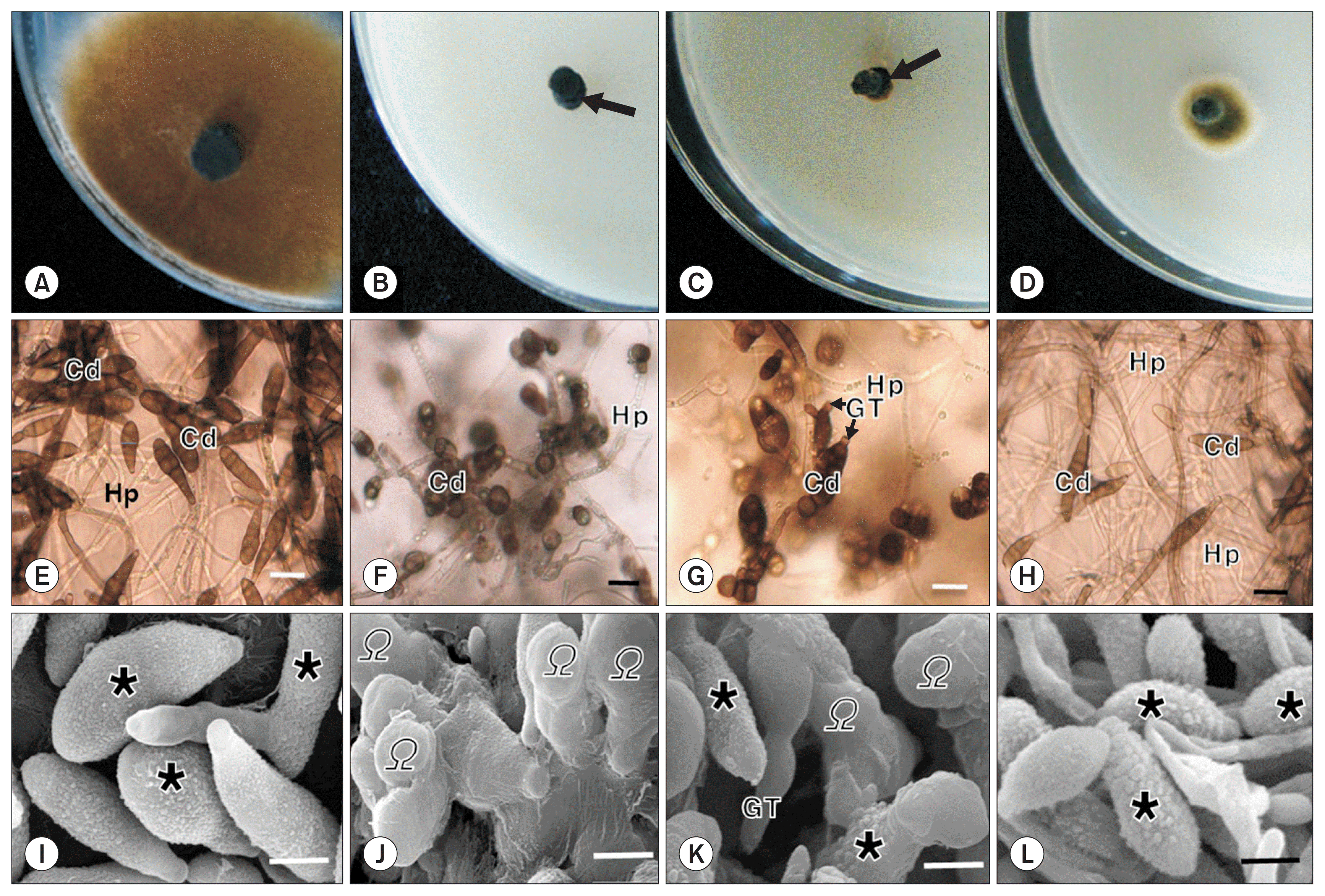
The fungal specimens for microscopic observations included four areas of 5-day-old fungal cultures on PDA with or without the fungicidal treatment, which were 1) fully grown culture (Con-G) in the untreated control, and three from the iprodione treatment, including 2) no growth (dead) (Ipr-N), 3) initially growing (living) (Ipr-I), and 4) fully grown fungal cultures (Ipr-G) (Fig. 2A–D). LM of Con-G and Ipr-G showed multicellular club-shaped (long ellipsoidal) dictyospores of 3–6 cross septa and 0–2 longitudinal walls with no morphological aberrations; severe conidial morphological aberrations occurred in Ipr-N, showing most of the conidia with spherical shapes with rare cross septa (Fig. 2E, F, H). For Ipr-I, unicellular spherical as well as multicellular club-shaped conidia were formed, accompanied by the occasional germination of the conidia indicated by outward protrusions (germ tubes) (Fig. 2G). SEM revealed conidial morphologies corresponding to their light microscopic features; long-ellipsoidal (club-shaped) conidia for Con-G and Ipr-G, mostly spherical shapes for Ipr-N, and mixture of club-shaped and spherical conidia with germ tubes for Ipr-I, respectively (Fig. 2I–L). Wart-like swellings were formed mostly on the surface of club-shaped (long-ellipsoidal) conidia, showing rough conidial surfaces (Fig. 2I, K, L), but rarely on the surface of spherical conidia, showing smooth conidial surfaces, especially on dead-looking conidia in Ipr-N (Fig. 2J).

Growth of Alternaria alternata on untreated potato-dextrose agar (PDA) (A) and PDA supplemented with 0.1% iprodione (B–D) after 5 days of incubation, showing full fungal growth in untreated control (A), non-growth (arrow; B), initial growth (arrow; C), full growth after one day after the initial growth (D) on PDA with iprodione and light (E–H) and scanning electron micrographs (I–L) of the fungal cultures corresponding to upper figures (A: E, I; B: F, J; C: G, K; D: H, L), showing conidia (Cd) and hyphae (Hp). GT, germ tube; asterisk (*), rough conidial surface with wart-like swellings mostly on ellipsoidal conidia; omega (Ω), smooth conidial surface with no wart-like swellings on spherical conidia. Scale bars = 20 μm (for light micrographs) and 10 μm (for scanning electron micrographs).
TEM
TEM of Con-G grown on PDA for 5 days without fungicide had conidia with cell walls and multi-lamellar bodies and dense, but intact, cytoplasm sometimes with cross walls (septa) (Fig. 3A, B). With no observable fungal growth (Ipr-N) on PDA with iprodione, most of the conidia shown were single-celled with a thick cell wall, and contained degenerated or extremely dense cytoplasm and occasional electron dense vacuolar inclusions and necrotic cytoplasmic contents (Fig. 3C–E). In the initial fungal growth on PDA with iprodione (Ipr-I), multicellular conidia with multiple septa were formed, and contained dense cytoplasm and proliferated lipid bodies; most conidia had two layers of outer (original) and inner (secondary) cell walls (Fig. 3F–H). In the mature colonies (Ipr-G) growing on PDA with iprodione, the conidial features were similar to those of the control (Con-G) with mono-layer of cell wall and dense intact cytoplasm sometimes containing occasional lipid bodies and prominent cell organelles such as mitochondria (Fig. 3I, J).

Transmission electron micrographs of Alternaria alternata on untreated potato-dextrose agar (PDA) (with full fungal growth) (A, B) and PDA supplemented with 0.1% iprodione (C–J) after 5 days of incubation with non-growth (C–E), initial growth (F–H), and full growth (I, J), showing ultrastructural features of conidia. Arrows, wart-like swellings. W, cell wall; Sp, septum; M, mitochondria; MB, multilamellar body; L, lipid body; V, vacuole; VI, vacuolar inclusion body; Nc, necrotic cytoplasmic contents; OW, outer cell wall; IW, inner cell wall; N, nucleus. Scale bars = 1.0 μm (A–D, F, G, I, J) and 0.5 μm (E, H).
Although no fungal growth occurred for the first few days on media supplemented with iprodione, it initiated and continued with increased frequencies with time. Once the initial growth (Ipr-I) began, the colonies grew and expanded continuously as in the untreated control, while no new mycelial growth initiated from no observable fungal growth region (Ipr-N) until the end of the experiment when the colonies of the initial growth grew fully with a sufficient sporulation. This suggests the initial fungal growth under fungicidal stress may be a turning moment, leading to further continuous development and growth. This indicates the morphogenetic changes at the initial growth on PDA with fungicide should be critical morphogenetic changes resisting the fungicidal toxicity.
In LM and electron microscopy (EM), all of the specimens with the same growth characteristics showed identical morphogenetic features of sporulation. When growing on media supplemented with iprodione, fungal sporulation was inhibited as illustrated by the microscopic features of mostly undeveloped unicellular conidia (indicating lack of cell division) with degraded cytoplasm sometimes containing necrotic cytoplasmic contents. The fungal colonies that initiated their growth in spite of the fungicide showed conspicuous morphogenetic alterations by forming multicellular conidia with thickened double layers of original outer cell wall and secondary inner cell wall. Fungal cell walls are rigid and structurally strengthened with complex polysaccharides, i.e., chitin and glucans, to provide structural barrier and to protect against osmotic lysis by acting as a molecular sieve and regulating the passage of large molecules through the wall (Deacon, 2006). Thus, the secondary inner conidial cell wall formed during the fungicidal treatment could decrease the penetration of the fungicide into conidial cells, and reduce fungicide-associated toxicity.
Another conspicuous cellular change during conidial formation in responses to the fungicidal stress was the accumulation of proliferated lipid bodies in the conidial cytoplasm. This response may be a means of counteracting one of the major fungicidal action modes of iprodione, inhibition of lipid metabolism (Fritz et al., 1977; Pappas and Fisher, 1979; Steel and Nair, 1995). In fungi, carbon is stored predominantly as triacylglycerol in lipid bodies in conidia that are used to sustain anabolism during conidial germination (Bago et al., 2000; Bonfante et al., 1994). Therefore, the growth of A. alternata in the presence of iprodione may be sustained by using energy and nutritional substances from lipid bodies protected by thickened inner and outer conidial cell walls. Collectively these results suggest that the resistant response begin at sporulation as exemplified by the morphogenesis of conidia with thickened cell walls and proliferated lipid bodies. More detailed examination of the interrelations of cell wall thickening and lipid body accumulation and their relations to oxidative protective mechanisms (free radical scavenging and catalase activity) (Steel and Nair, 1995) would provide better understandings on the fungicide detoxification mechanisms of the fungus, which may be useful for the development of strategies for the practical control of the Alternaria diseases.
