Characterization of the rcsA Gene from Pantoea sp. Strain PPE7 and Its Influence on Extracellular Polysaccharide Production and Virulence on Pleurotus eryngii
Article information
Abstract
RcsA is a positive activator of extracellular polysaccharide (EPS) synthesis in the Enterobacteriaceae. The rcsA gene of the soft rot pathogen Pantoea sp. strain PPE7 in Pleurotus eryngii was cloned by PCR amplification, and its role in EPS synthesis and virulence was investigated. The RcsA protein contains 3 highly conserved domains, and the C-terminal end of the open reading frame shared significant amino acid homology to the helix-turn-helix DNA binding motif of bacterial activator proteins. The inactivation of rcsA by insertional mutagenesis created mutants that had decreased production of EPS compared to the wild-type strain and abolished the virulence of Pantoea sp. strain PPE7 in P. eryngii. The Pantoea sp. strain PPE7 rcsA gene was shown to strongly affect the formation of the disease symptoms of a mushroom pathogen and to act as the virulence factor to cause soft rot disease in P. eryngii.
Introduction
Pleurotus eryngii was originally cultivated in northern Italy and Switzerland, where it is known locally as cardoncello (Ohga and Royse, 2004). But it is now commonly cultivated in Europe, the Middle East, and North America, as well as in parts of Asia. Cultivation of P. eryngii on an industrial scale began in Korea in 1996, and by 2014, production of P. eryngii was estimated to be 47,814 tons (Ministry for Food, Agriculture, Forestry and Fisheries, 2015). It is susceptible to external conditions such as temperature, humidity, and carbon dioxide (CO2) levels. As a result of this, it is more prone to disease and more sensitive to other growing conditions. To date, it has been reported a number of fungal and bacterial diseases such as dry bubble disease on Agaricus bitorquis by Verticillium fungicola var. fungicola, internal stipe necrosis on A. bitorquis by Ewingella americana, and bacterial soft rot and black rot on Flammulina velutipes by Erwinia carotovora subsp. carotovora and Pseudomonas tolaasii (Gea et al., 2003; Han et al., 2012; Inglis et al., 1996; Okamoto et al., 1999). They are involved in the major steps of the cultivation process. Especially, blotch diseases have been frequently reported in cultivated mushrooms. These are mainly due to P. tolaasii, causing brown blotch disease, and Pseudomonas gingeri, responsible for ginger blotch disease (Flippi et al., 2002; Paine, 1919; Tolass, 1915; Wong et al., 1982). Wells et al. (1996) described a mild light-brown discoloration with slight tissue collapse in the cultivated mushroom Agaricus bisporus that was attributed to Pseudomonas reactans another common inhabitant of the mushroom hyphosphere. The gram negative bacterium Pantoea sp. causes soft rot disease with symptoms of water-soaked lesions on the stipes and pileus of P. eryngii (Kim et al., 2007). The symptoms of the disease also include a dark-brown drop that developed on hypha and primordia of the mushrooms in the early stage. Water soaked lesions had developed on the stipes and pileus, and the normal growth of the mushrooms was inhibited. An offensive odor then developed along with a severe soft rot, which can result in serious economic damage. The genus Pantoea was established in 1989 and includes plant pathogenic species formerly classified as Erwinia herbicola, E. uredovora, E. milletiae, E. ananas, and E. stewartii (Gavini et al., 1989; Mergaert et al., 1993). Pantoea ananatis was first reported as a pathogen of pineapple fruit, causing brown rot (Serrano, 1928). Since then, it has been reported to cause disease on sudangrass, cantaloupe fruit, onion, eucalyptus species, and honeydew melons (Azad et al., 2000; Bruton et al., 1991; Ceponis et al., 1985; Coutinho et al., 2002; Gitaitis and Gay, 1997). Pantoea stewartii (formerly Erwinia stewartii) is a vascular wilt and leaf blight pathogen of corn whose primary mechanism of virulence is production of extracellular polysaccharides (EPSs) slime that occludes the xylem vessels of the plant (Braun, 1982; Gea et al., 2003; Mergaert et al., 1993). Polysaccharides can remain associated with the cell wall to form a bound capsule layer, or they can be released into the cell’s milieu as extracellular slime. Most pathogenic and symbiotic bacteria are EPS. The production of EPS is a common feature of many gram negative bacteria, especially plant pathogens (Leigh and Coplin, 1992; Sutherland, 1985). EPS creates a favorable microenvironment for the bacterial cells and protects them against desiccation, external stress, and host defense mechanisms (Bereswill and Geider, 1997; Dolph et al., 1988). EPS is furthermore an essential determinant for the bacterial virulence in several host-pathogen interactions such as Erwinia amylovora and P. stewartii subsp. stewartii (Leigh and Coplin, 1992). The structure of EPS is highly variable, and different types have been classified by molecular weight and structural properties (Jayaratne et al., 1993). Many of these polysaccharides are acidic heteropolysaccharides that have repeating subunit structures with carbohydrates that contain neutral sugars and uronic acids and noncarbohydrate substituents. Others are homopolysaccharides such as levan in Erwinia and Pseudomonas species, alginate in Azotobacter and fluorescent Pseudomonas species, cellulose in Agrobacterium and Rhizobium species, and glucan in Rhizobiaceae sp. (Bennett and Billing, 1980; Fett et al., 1989; Fyfe and Govan, 1980; Hisamatsu et al., 1987; Matthysse, 1983; Smit et al., 1987). The biosynthesis of the high molecular weight EPS in several enterobacterial species is modulated by the regulation of capsule synthesis (Rcs) regulatory network (Gottesman, 1995). The nucleotide sequence of the Klebsiella pneumoniae rcsA gene was the first to be determined by Allen et al. (1987) so that the subsequently reported sequences of E. amylovora rcsA and Escherichia coli rcsA were individually compared to it (Bernhard et al., 1990; Coleman et al., 1990; Stout et al., 1991). These RcsA proteins had significant homology with the C-terminal and N-terminal ends. In the case of E. coli, capsular polysaccharide synthesis is encoded by the cps genes and is under the control of at least 2 positive regulators, the products of the rcsA and rcsB genes (Brill et al., 1988; Gottesman et al., 1985; Stout and Gottesman, 1990; Torres-Cabassa et al., 1987). Torres-Cabassa et al. (1987) showed that the cpsA–cpsD genes in E. stewartii are regulated by the rcsA gene that is functionally equivalent to rcsA in E. coli. The inactivation of rcsA caused a strong reduction of EPS synthesis that was associated with reduced virulence in E. stewartii. Although E. amylovora rcsA does not share extensive similarity with the other rcsA gene, disruption of the gene showed a significant decrease in amylovoran production and reduced virulence (Chatterjee et al., 1990; Coleman et al., 1990). This study was conducted to identify and characterize of the pathogenicity gene from the soft rot pathogen Pantoea sp. PPE7 in P. eryngii. The nucleotide and amino acid sequences of rcsA, an open reading frame (ORF) that was identified from Pantoea sp. strain PPE7, were characterized by sequence analysis. In addition, a Pantoea sp. strain PPE7 null mutant in which the rcsA gene was inactivated was constructed by a gene replacement strategy and confirmed that the RcsA protein was involved in the development of soft rot disease with symptoms of water-soaked lesions in P. eryngii.
Materials and Methods
Bacterial strains, plasmids, and culture conditions
The bacterial strains and plasmids used in this study are listed in Table 1. The Pantoea sp. strain PPE7 was grown at 28°C in nutrient broth or Luria-Bertani (LB) broth containing 10 g of Bacto-tryptone, 5 g of yeast extract, and 5 g of NaCl per liter of medium (pH 7.0). E. coli strains containing the recombinant plasmid were grown at 37°C in LB medium or LB medium supplemented with the appropriate antibiotics. All antibiotics were purchased from Sigma and were used at the following concentrations: ampicillin 50 μg ml−1, kanamycin 50 μg ml−1. The culture media were purchased from Difco (Detroit, MI, USA).

Bacterial strains and plasmids used in this study
Substrate preparation and cultivation methods for P. eryngii.
The substrate for the growth and fruiting of P. eryngii consisted of 55% sawdust from the pine tree and poplar (mixing ratio, 2:1), 25% wheat bran, and 20% rice bran (v/v, in terms of dry weight). The final moisture content of the substrate was 65%. Approximately 550 g of substrate was packed into 850 ml polypropylene bottles and sterilized at 121°C for 90 min. After autoclaving, bottles were cooled at 20°C in a cooling room. Inoculated bottles with the spawn were moved to an incubation room where the temperature and humidity were maintained at 22°C to 24°C and 65% to 68%, respectively, and incubated for 35 days. When the substrate was colonized, the spawn was removed and water was added to the media in the bottles. The bottles were then transferred to a cultivation room for 20 days to obtain fruiting bodies. Fruiting was induced by maintaining a low temperature (about 15°C) and high humidity (approximately 90% to 95%).
Construction of Pantoea sp. strain PPE7 expressing the green fluorescent protein
The pGFP with the lacZ initiation codon from pUC19 was used for tagging the Pantoea sp. strain PPE7 with the gfp gene expressing the green fluorescent protein. Pantoea sp. strain was transfected by electroporation and the transfected cells were selected on LB agar containing ampicillin (50 μg ml−1). Green fluorescent colonies were visualized by using a portable UV lamp (UVGL-58; UVP, Upland, CA, USA). The strain expressing green fluorescent protein was named Pantoea sp. PPEG7.
DNA methods and sequence analysis
Genomic DNA from Pantoea sp. strain PPE7 was isolated by the method described by Ausubel et al. (1999). Plasmid DNA was isolated by an alkaline method or DNA-spinTM Plasmid DNA Extraction Kit (iNtRON Biotechnology, Suwon, Korea). Restriction enzymes and DNA-modifying enzymes were purchased from iNtRON (iNtRON Biotechnology) and Promega (Madison, WI, USA). Other chemicals were purchased from Sigma Chemical Co. (St. Louis, MO, USA). Computer-assisted sequence analyses were performed utilizing the DNAMAN analysis system (Lynnon Biosoft, Quebec, Canada). DNA and amino acid sequence homology searches were done using the National Center for Biotechnology Information (http://www.ncbi.nlm.nih.gov) at the National Library of Medicine (Bethesda, MD, USA). SOSUI and PROSITE searches were performed via the ExPASy World Wide Web molecular biology server (http://www.expasy.org) from the Geneva University Hospital and the University of Geneva, Switzerland.
Electrotransformation of Pantoea cells
Competent Pantoea cells were prepared for electrotransformation in 10% glycerol. Electroporation was performed with a Gene-Pulser (Bio-Rad, Hercules, CA, USA) using 80 μl of an electrocompetent Pantoea cell suspension and 10 to 200 ng of plasmid DNA at 2.5 kV, 25 F, and 200 Ω. Two minutes after electroporation, the cell suspension was diluted in 800 μl of LB broth and incubated for 1 h at 28°C with shaking; this was followed by plating on selective medium.
Pathogenicity tests
Bacterial suspensions were prepared from 24 h LB broth cultures to inoculate the P. eryngii. When the substrate was colonized by the mycelium of mushroom, the spawn was removed and inoculated with 10 ml of bacterial suspensions (approximately 1 × 10⁶ cfu ml−1). Inoculated bottles were incubated in a cultivation room at 16°C to 17°C. The symptoms of disease development were examined for 20 days. Once symptoms were expressed, several pieces with lesions from stipes and pileus were crushed with a sterile pestle, and suspended with 10 ml of distilled water. Bacteria were re-isolated on nutrient agar medium. The re-isolated strains were compared with the inoculated strain using 16S rDNA sequence analysis. The experiment was repeated at least three times.
Construction of the disrupted rcsA gene by random insertion mutagenesis
To construct pRCS101 with a transposon that inserted into the rcsA gene, we used an EZ-Tn5 <KAN-2> insertion as suggested by the manufacturer (Epicentre, Madison, WI, USA). The 1.2-kb EZ-Tn5 element, derived from Tn903, carries a kanamycin (Km) resistance gene flanked by an inverted repeat sequence. Electroporation was performed with a Gene-Pulser (Bio-Rad) using 80 μl of an electrocompetent DH5α cell suspension and 100 ng of plasmid DNA at 2.5 kV, 25 F, and 200 Ω. Transposition clones were selected by plating on LB agar medium containing ampicillin (50 μg ml−1) and kanamycin (50 μg ml−1). Transposon insertion sites were mapped by PCR and automated sequencing using the following specific primers: forward primer KAN-2-FP-1 (5′-ACCTACAACAAAGCTCTCATCAACC-3′) and reverse primer KAN-2-RP-1 (5′-GCAATGTAACATCAGAGATTTTGAG-3′).
Construction of the rcsA mutant strain, Pantoea sp. NPPE148
The disrupted rcsA gene fragment of plasmid pRCS101 was cloned into the suicide vector pEX18Ap, resulting in pRCSEX101. The suicide vector pEX18Ap carries an ampicillin resistance determinant and sacB, the levan sucrase-encoding gene, which confers sucrose sensitivity in E. coli strains when grown on LB medium supplemented with 5% sucrose (Hoang et al., 1998). This construct was transformed by electroporation into Pantoea sp. strain PPE7 electrocompetent cells. Cells were plated on LB agar containing kanamycin (50 μg ml−1) to select Pantoea sp. strain PPE7 transformants. Subsequently, colonies that grew on LB agar containing kanamycin (100 μg ml−1) plates were streaked on LB containing 10% sucrose. This step was repeated 2 times to apply selective pressure to merodiploids to prevent their growth on sucrose-containing LB medium. Colonies from sucrose selection medium were inoculated onto both LB agar containing kanamycin (50 μg ml−1) and ampicillin (50 μg ml−1). The colonies, which exhibited a kanamycin-resistant, ampicillin-sensitive phenotype, were screened by PCR using the following specific primers: forward primer GARES11 (5′-CAGCCAACGAATCCGTAG-3′) and reverse primer GARES12 (5′-CATTGTCCGTTAATCGCA-3′). The strain was designated Pantoea sp. NPPE148 and contained the disrupted rcsA gene.
Construction and analysis of cosmid library
The cosmid library of Pantoea sp. strain PPE148 genomic DNA was constructed in pCC1FOSTM according to the protocol recommended by the manufacturer (CCFOS110; Epicnetre, Madison, WI, USA). The library was replicated into duplicate sets in 1.5 ml micro-tubes and stored individually in 80% glycerol at −20°C. The diversity and average size of cosmid library were estimated by agarose gel electrophoresis of EcoRI and BamHI digested fragments. The average insert size of the cosmid clones was approximately 40 kb, ranging from 9–49 kb. The cosmid clone, which contained the rcsA gene, was screened by PCR using the following specific primers: forward primer GARES11 (5′-CAGCCAACGAATCCGTAG-3′) and reverse primer GARES12 (5′-CATTGTCCGTTAATCGCA-3′). The resulting cosmid clone was designated pPCOS429.
Construction of pRCS100, 200, and 300 for complementation
For the complementation experiments, we reconstructed the 2.2 kb intact rcsA gene in the pGEM-T easy vector by PCR using the following specific primers: forward primer GARES 77 (5′-CTCGGTCTGGTAATGCCT-3′) and reverse primer GARES 78 (5′-ATGTTAAGAGCAGGGCAG-3′). The resulting plasmid was designated pRCS100. The rcsA gene of Pantoea sp. strain PPE7 was isolated by digesting pRCS100 with EcoRI restriction enzyme and was cloned into pBluescript II SK+ and the pACYC184-based vector pSTV28 to yield the complementation constructs pRCS200 and pRCS300. The resulting plasmid contained the rcsA gene in the same orientation as the lac promoter, and it was transformed by electroporation into Pantoea sp. NPPE148 electrocompetent cells. The transformants were selected on LB agar containing kanamycin (50 μg ml−1) and chloramphenicol (30 μg ml−1). The virulence of strains was examined by a pathogenicity test.
EPS determination
EPS was extracted from a bacterial suspension on minimal agar plate for 24 h (Torres-Cabassa et al., 1987). Bacteria were scraped from the plate and suspended to yield A540 = 0.5 (1 × 10⁻⁸ cfu ml−1) in PBS buffer (10 mM potassium phosphate buffer, pH 7.0, 15 mM NaCl, 1 mM MgSO4·7H2O), and the cells were removed by successive centrifugation at 3,000g for 30 min and 16,000g for 60 min at 4°C. EPS was precipitated with 3 volumes of 95% ethanol overnight at −20°C and then was redissolved in distilled water. Total carbohydrates, based on glucose, were determined according to the anthrone method with the following modification (Coplin et al., 1986). The anthrone reagent was prepared by dissolving 0.1 g of anthrone in a solution of 100 ml of H2SO4 (specific gravity 1.84), and 33 ml of distilled water was added. One milliliter samples of ethanol extract that were obtained from bacterial suspension were mixed with 5 ml of anthrone reagent, heated at 100°C for 10 min, and read immediately at A625.
Confocal microscope analysis
For the in situ microscopic analysis of the specimens, bacterial suspensions (Pantoea sp. PPEG7, approximately 1.0 × 10⁶ cfu ml−1) were prepared from 24 h LB broth cultures to inoculate the mycelium of P. eryngii cultivated for 7 days in a 250 ml Erlenmeyer flask. The mycelium was then washed 3 times by shaking for 5 min in distilled water. These speciman were mounted on a slide glass and visualized using a confocal laser scanning microscope (FV1000; Olympus, Tokyo, Japan). For the confocal microscopy the excitation wavelength was 488 nm (Argon ion laser) and the emitted light was detected in the range of 510–560 nm.
Examination of infectious fruitbody by scanning electron microscope
For scanning electron microscope (SEM), 10 ml of bacterial suspensions (Pantoea sp. NPPE148 [pRCS300], approximately 1.0 × 10⁶ cfu ml−1) was prepared from 24 h LB broth cultures to inoculate the mycelium of P. eryngii. When symptoms were expressed, pieces of stipes from the diseased mushrooms were fixed in 5% glutaraldehyde in sodium phosphate buffer (pH 7.3) for 2 h at room temperature. The preparations were washed 3 times in the buffer and then were metallized using osmium tetroxide and thiocarbohydrazide. This was followed by dehydration in ethanol and Freon 113 before critical point drying. To assure surface conductance, the samples were coated with a 300-Å gold layer by high vacuum evaporation using a rotating stage. The specimens were then examined with a model VT1420 (Carl Zeiss, Oberkochen, Germany) SEM using a beam accelerating voltage of 25 kV.
Statistical analysis
All data were statistically analyzed using the SAS software version 9.2 (SAS Institute, Cary, NC, USA). The measured EPS contents were compared using a one-way ANOVA. In addition, the significant effect in EPS contents was detected, and the Bonferroni t-test was used to detect and separate the mean treatment differences at 5.0% levels of significance (P < 0.05).
Results and Discussion
Interaction of Pantoea sp. PPEG7 with P. eryngii
When the substrate in the bottles was colonized, the spawn was removed by a scratching method and simultaneously inoculated with Pantoea sp. PPEG7 to investigate the interaction between P. eryngii and the pathogen. After 5 to 7 days, the symptoms of the disease, which include a dark-brown water droplet, developed on the hypha (Fig. 1A) and primordium (Fig. 1B) of the mushrooms inoculated with the strain expressing green fluorescent protein and inhibited the normal growth of fruit body (Fig. 1C). Green fluorescent colonies were visualized in internal parts of the infected fruitbody under UV light. The mycelium of P. eryngii inoculated with a suspension of the strain expressing green fluorescent protein was directly visualized by confocal microscopy (Fig. 1D). The bacteria could be easily observed on the surface of the mycelium because of the strong fluorescence signals in the cells. Green fluorescent bacteria formed a virtually continuous layer of tightly aggregated cells around the mycelium and clamp connection of P. eryngii. These results showed that Pantoea sp. strains PPE7 could affect the mycelial growth of P. eryngii by direct interaction and provide a major factor for the disease appearance.
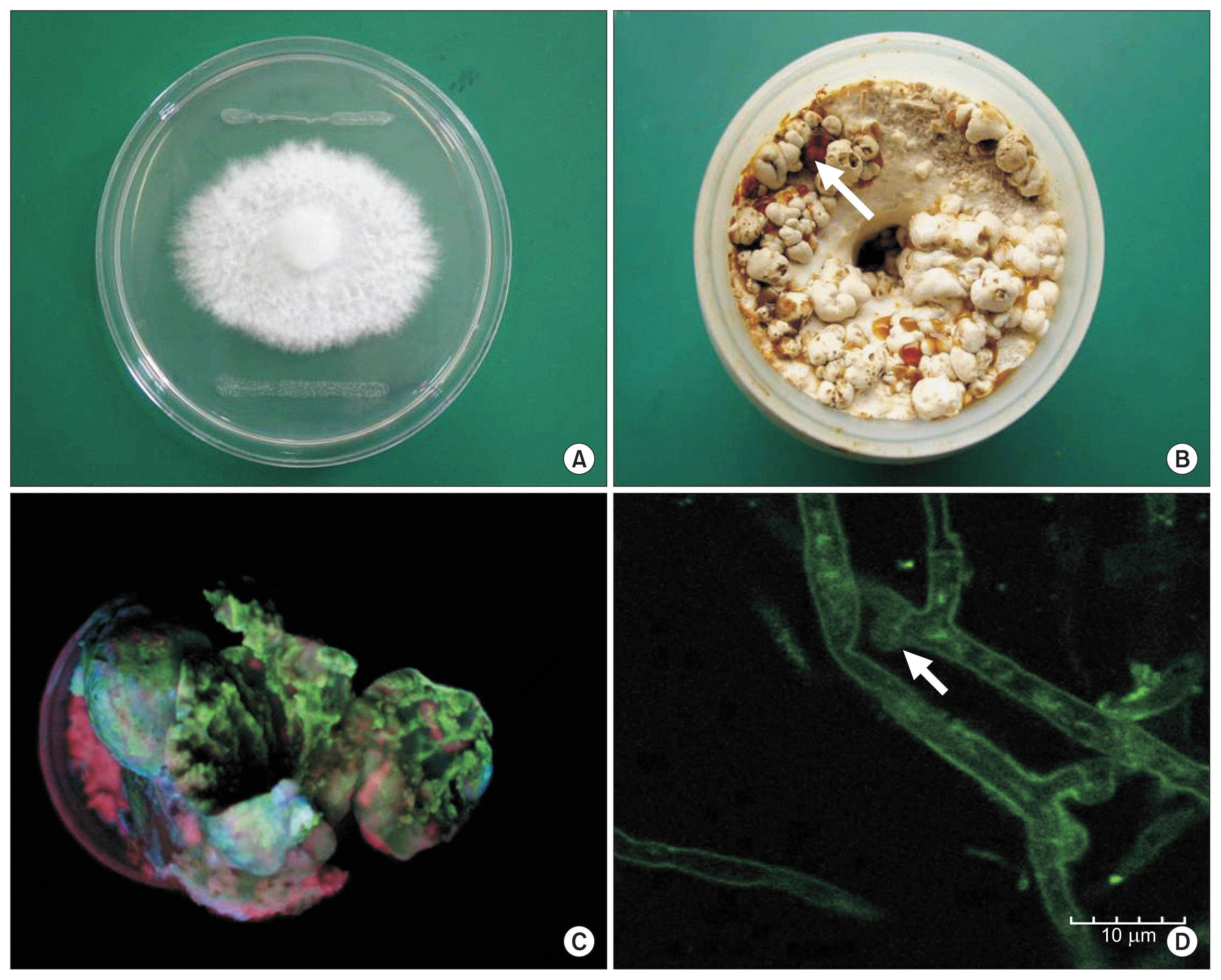
Symptoms of soft rot disease (A–C) of Pleurotus eryngii and confocal microscopic photographs (D) of mycelium after artificial inoculation with Pantoea sp. PPEG7 expressing green fluorescent protein. Inoculation of pathogen induced a dark brown water drop in early stage and developed viscous fluid on fruitbody. Visible (B) and fluorescent (C) photograph of the inoculated mushroom were recorded at 7 and 12 days postinoculation. (A) Mycelium inhibited by Pantoea sp. PPEG7. (B) Primordial. (C) Fruitbody. (D) Mycelium. The arrows indicate a viscous fluid (B) on the stage of primordia formation and the clamp connection (D) of P. eryngii.
Sequence and identification of the Pantoea sp. strain PPE7 rcsA gene
To construct a small clone that carried a functional Pantoea sp. PPE7 rcsA, a 2.2-kb fragment was amplified by PCR and cloned into the pGEM-T easy vector to produce plasmid pRCS100. The nucleotide sequence and deduced amino acid sequence of the rcsA gene are shown in Fig. 2. Two possible ORFs were present in the sequence of pRCS100, and both started with ATG codons at positions +1 and +16. The ATG at +1 was designated as the start codon because a putative ribosome binding site (GAGG) was present 10 bp upstream and because sequence similarity with the E. coli RcsA protein, the amino acid sequence of which has been determined, also begins at this position (Poetter and Coplin, 1991; Stout et al., 1991). The rcsA gene consists of a 633 bp coding region and encodes a predicted protein of 211 amino acids. The molecular mass of the RcsA protein is about 25 kDa. The sequence of the rcsA gene has been assigned GenBank accession number KC537802.

Comparison of the genomic organization based on the physical map of cosmid pPCOS429 carrying the rcsA gene (A) and nucleotide and deduced amino acid sequence (B) of rcsA of Pantoea sp. PPE7. A putative ribosomal binding site (GAGG) and a possible Escherichia coli-10 consensus promotor sequence (ATAA) are marked with single underlines. The start (ATG) and stop (TAA) codon are marked with a bold characters. Position of the inserted kanamycin gene is indicated by an inverted triangle and double underline.
Comparison and analysis of known RcsA proteins
In previous studies, the amino acid sequences of E. amylovora RcsA and E. coli RcsA were compared with that of K. pneumoniae RcsA (Allen et al., 1987; Bernhard et al., 1990; Coleman et al., 1990; Stout et al., 1991). The following is a comparison of all 5 known RcsA amino acid sequences (Fig. 3). The RcsA protein contains 3 highly conserved domains. The first 20 amino acids are highly conserved in all species. In this region, approximately 65.0% of all nucleotide sites are invariant, and 65.0% of the polymorphic sites are in the third position of a codon. The amino acid similarities are likewise pronounced in this region of the protein. The second region, amino acid positions 50 to 60, is almost invariant. The few amino acid substitutions in this region are all conservative: a phenylalanine to isoleucine replacement in E. amylovora, aspartate to glutamate replacement in Pantoea sp. strain PPE7, and isoleucine to leucine and isoleucine to valine substitutions in K. pneumoniae. The third and most striking region of similarity in the gene extends from amino acids 145 to 187. The degree of amino acid conservation in this region is 76.0% identity in all of the pairwise comparisons. The alignment of amino acid sequences suggested that RcsA may have the properties of a DNA-binding protein and be involved in regulating EPS synthesis by directly activating polysaccharide synthesis promoters. Torres-Cabassa et al. (1987) showed that the cpsA–cpsD genes in E. stewartii are regulated by an rcsA gene that is functionally equivalent to rcsA in E. coli. Computer analysis of the RcsA proteins revealed that a helix-turn-helix motif existed in the C-terminal region. This result was accordance with that suggested by Pabo and Sauer (1984).

Comparative analysis of the predicted amino acids of Pantoea sp. strain PPE7 RcsA homologous. P. sp, Pantoea sp. strain PPE7; P. s, Pantoea stewartii subsp. stewartii; E. a, Erwinia amylovora; E. c, Escherichia coli; K. p, Klebsiella pneumoniae; S. t, Salmonella typhimurium LT2. The boxes indicate three highly conserved domains in the RcsA amino acids. Asterisks indicate identical amino acid, and dots indicate similar amino acid.
Analysis and characterization of the rcsA mutant strain, Pantoea sp. NPPE148
To investigate the function of the rcsA gene, it was disrupted by the insertion of the kanamycin-resistance cassette of EZ-Tn5 transposon using pRCSEX101. An rcsA::Kmr mutant, Pantoea sp. NPPE148, was generated by a gene replacement strategy as previously described by Hoang et al. (1998). The insertion of the 1.2 kb kanamycin-resistance cassette was confirmed by PCR analysis using the rcsA specific primers GARES11 and GARES12. The 1.6 kb PCR product obtained from the mutant indicated the correct replacement of pRCSEX101 into the intact rcsA gene. To analyze the rcsA gene and the flanking region in Pantoea sp. strain PPE148, the cosmid clone containing the kanamycin gene in mutant Pantoea sp. NPPE148 was constructed. The cloned genomic DNA was initially analyzed by determining the nucleotide sequence on either side of the kanamycin gene insertion point by using primers specific for either end of the kanamycin gene. The flanking region of an rcsA gene into which the kanamycin gene had been inserted was determined using the cosmid clone pPCOS429 and internal oligonucleotide primers (Fig. 2A). The site of insertion of the kanamycin gene in mutant Pantoea sp. NPPE148 was found to be located between position 416 and 417 of the coding region of the rcsA gene (Fig. 2B). A comparison of genomic organization showed that the rcsA gene of Pantoea sp. PPE7 have the same transcriptional direction and highly homologous to other strains. Pantoea sp. NPPE148 containing the disrupted rcsA gene showed non-mucoid, non-convex and irregular colony morphology. The virulence of Pantoea sp. strains PPE7 and NPPE148 was analyzed on the mycelium of P. eryngii that was cultivated for 35 days in bottles. Inactivating rcsA abolished the virulence of Pantoea sp. strain PPE7 in P. eryngii (Fig. 4D, E) whereas the wild-type strain displayed droplets of ooze and water-soaked lesions on mycelium and primordial after 10 days (Fig. 4B, C).
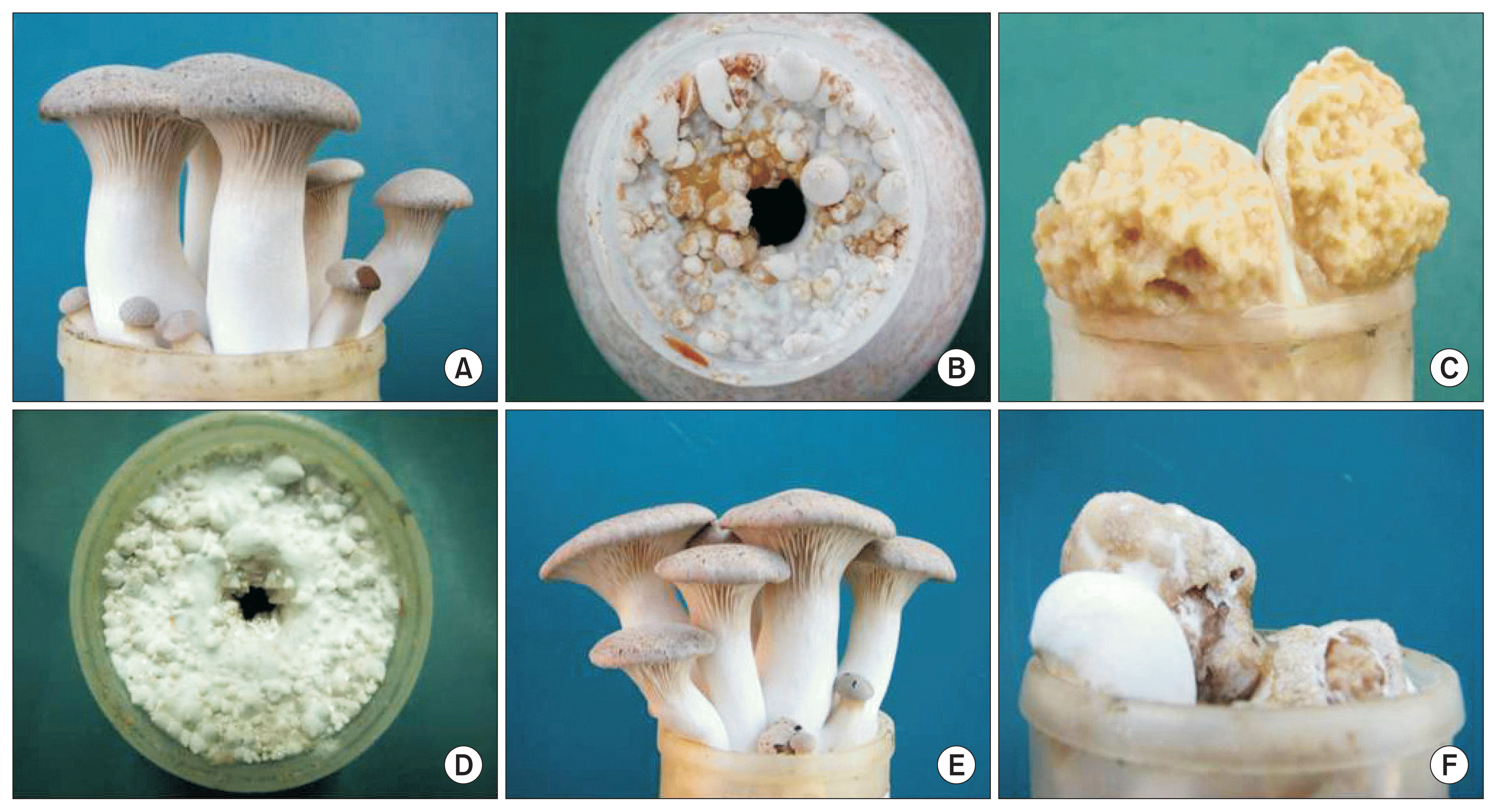
Pathogenicity test for the derivatives of Pantoea sp. strain PPE7. Bacterial suspensions (approximately 1.0 × 10⁶ cfu ml−1) were prepared from 24 h Luria-Bertani broth cultures to inoculate the mycelium of Pleurotus eryngii cultivated for 35 days in bottles. (A) Control. (B, C) Pantoea sp. strain PPE7. (D, E) Pantoea sp. NPPE148. (F) Pantoea sp. NPPE148 harboring pRCS300.
Complementation of Pantoea sp. NPPE148 and the influence of rcsA on virulence
To determine the role of rcsA in pathogenicity, various plasmids with an intact rcsA gene were introduced into the mutant strain, Pantoea sp. NPPE148. The colony morphology analysis showed that Pantoea sp. NPPE148 containing pRCS100 and pRCS200 which are high-copy plasmids that carry a 2.2 kb intact rcsA gene region had a mucoid colony morphology indicative of increased EPS production (data not shown). A similar increase in EPS production was reported in E. coli cells containing multiple copies of rcsA, a positive regulator of EPS synthesis (Gottesman et al., 1985). However, these plasmids did not restore completely the pathogenicity of the mutant strain against P. eryngii. In contrast, the introduction of pRCS300, which is a low-copy plasmid carrying a 2.2 kb intact rcsA gene region, into Pantoea sp. NPPE148 resulted in the complete restoration of virulence against P. eryngii (Fig. 4F).
The restoration of virulence indicated that this mutant does not have a second-site mutation that is responsible for its pathogenicity and phenotype. These results indicated that the rcsA gene affects the pathogen’s major mechanism of virulence and requires low-level expression rather than high-level expression for virulence.
Detection of pathogens in the infected mushroom
To examine the adhesion of pathogens in an infected mushroom, we visualized the surface of the mushroom by SEM. The colonization and aggregation of Pantoea sp. PPE7 and Pantoea sp. NPPE148 on the surface of an infected mushroom is presented in Fig. 5. Pantoea sp. PPE7 was associated with hypha of mushroom. Mutants did not show the evidence of aggregation or of the production of a fibrillar matrix (Fig. 5D). Pantoea sp. NPPE148 was reduced in the population and cell-to-cell aggregation on the surface of the mushroom. Observation of Pantoea sp. NPPE148 on the surface revealed that cells were not presented in the intercellular spaces of hypha. Considering this observation, we assumed that the pathogen can grow on the surface of mycelium and intercellular spaces of mushrooms. The production of EPS is thought to contribute to the colonization and adhesion in the surface of mushroom by Pantoea sp. PPE7.
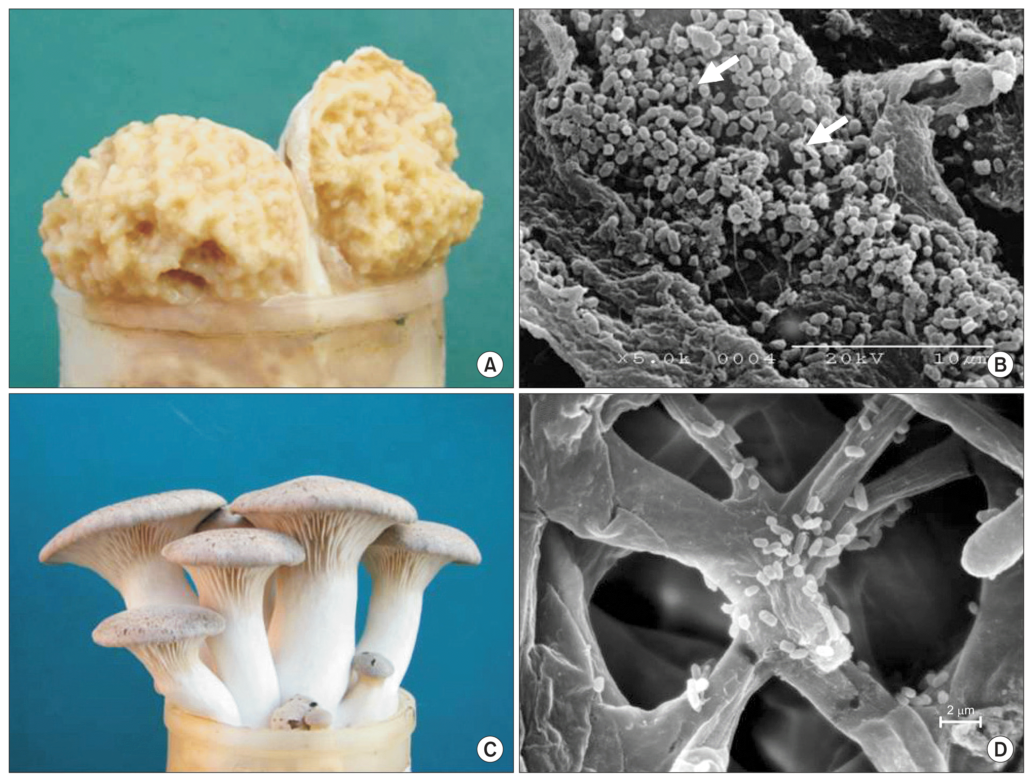
Symptoms of Pleurotus eryngii after inoculation with bacteria (A, Pantoea sp. PPE 7; C, Pantoea sp. NPPE148) and scanning electron micrographs (B, Pantoea sp. PPE7 5,000×; D, Pantoea sp. NPPE148 10,000×). Bacterial suspensions (approximately 1.0 × 10⁶ cfu ml−1) were prepared from 24 h Luria-Bertani broth cultures to inoculate the mycelium of Pleurotus eryngii cultivated for 35 days in bottles. The arrows indicate a fibrillar matrix on the surface of the mushroom.
Relationship between rcsA and EPS production
To determine whether the rcsA gene affects EPS synthesis, the EPS content was measured in a bacterial suspension by the anthrone method (Fig. 6). Pantoea sp. NPPE148 had decreased production of EPS (6.8 × 10⁻⁸ μg cell−1) compared to the wild-type strain (19.7 × 10⁻⁸ μg cell−1) and showed a nonmucoid colony morphology on minimal agar. The wild-type phenotype could be restored (22.7 × 10⁻⁸ μg cell−1) by complementation of Pantoea sp. NPPE148 with plasmid pRCS300. The colony showed a mucoid morphology indicative of increased EPS production and the complete restoration of virulence against P. eryngii. A similar increase in polysaccharide production was reported in E. coli and E. stewartii cells containing multiple copies of rcsA, a positive regulator of colanic acid synthesis (Chatterjee et al., 1990; Gottesman et al., 1985). These results supported the idea that EPS synthesis is affected by the product of the rcsA gene in Pantoea sp. PPE148 and rcsA gene may act as the virulence factor to cause soft rot disease in P. eryngii. These results led us to confirm that the rcsA gene in Pantoea sp. PPE7 was related to the pathogenicity and an important component that causes soft rot disease in P. eryngii. Also, these provide a better understanding of the soft rot disease by Pantoea sp. strain PPE7 in P. eryngii.

Influence of rcsA on extracellular polysaccharide (EPS) synthesis of various strains grown on minimal agar. EPS was extracted from bacterial suspension made from 24 h minimal agar plate cultures. The EPS contents based on glucose for cells suspended were determined by anthrone method for the wild-type strain Pantoea sp. PPE7, for Pantoea sp. NPPE148 with mutation in rcsA, and for Pantoea sp. NPPE148 (pRCS300) with Pantoea sp. NPPE148 harboring pRCS300. Sample means were compared by an analysis of variance and separated using Bonferroni t-test. The presence of different letters above sample mean values indicates that the means were significantly different at P < 0.05.
Acknowledgments
This work was carried out with the support of “Research Program for Agriculture Science and Technology Development” Gyeongsangnam-do Agricultural Research & Extension Services, Republic of Korea.